
solo per uso di ricerca
CUDC-101 HDAC inibitore
N. Cat.S1194
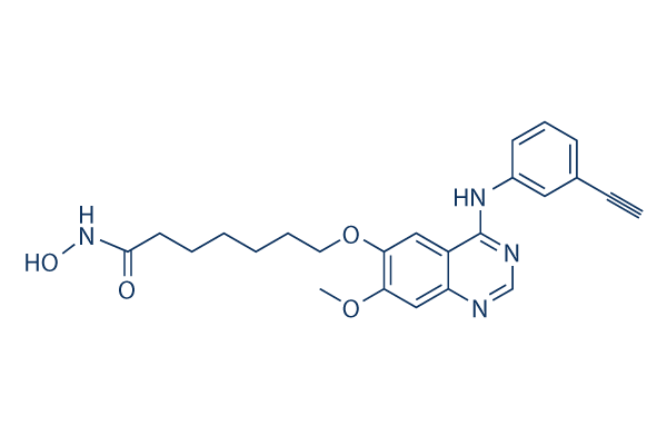
Struttura chimica
Peso molecolare: 434.49
Controllo Qualità
Coltura cellulare, trattamento e concentrazione di lavoro
| Linee cellulari | Tipo di saggio | Concentrazione | Tempo di incubazione | Formulazione | Descrizione dellattività | PMID |
|---|---|---|---|---|---|---|
| human SK-BR-3 cells | Proliferation assay | Antiproliferative activity against human SK-BR-3 cells after hrs by ATP content assay, IC50=0.04 μM | ||||
| MDA-MB-231 cells | Proliferation assay | Antiproliferative activity against human MDA-MB-231 cells after hrs by ATP content assay, IC50=0.1 μM | ||||
| human HepG2 cells | Proliferation assay | Antiproliferative activity against human HepG2 cells after hrs by ATP content assay, IC50=0.13 μM | ||||
| human SKHEP1 cells | Proliferation assay | Antiproliferative activity against human SKHEP1 cells after hrs by ATP content assay, IC50=0.22 μM | ||||
| human Hep3B2 cells | Proliferation assay | Antiproliferative activity against human Hep3B2 cells after hrs by ATP content assay, IC50=0.23 μM | ||||
| human BxPC3 cells | Proliferation assay | Antiproliferative activity against human BxPC3 cells after hrs by ATP content assay, IC50=0.27 μM | ||||
| human NCI-H358 cells | Proliferation assay | Antiproliferative activity against human NCI-H358 cells after hrs by ATP content assay, IC50=0.4 μM | ||||
| human MCF7 cells | Proliferation assay | Antiproliferative activity against human MCF7 cells after hrs by ATP content assay, IC50=0.55 μM | ||||
| human HCC827 cells | Proliferation assay | Antiproliferative activity against human HCC827 cells after hrs by ATP content assay, IC50=0.6 μM | ||||
| human H460 cells | Proliferation assay | Antiproliferative activity against human H460 cells after hrs by ATP content assay, IC50=0.7 μM | ||||
| human Capan1 cells | Proliferation assay | Antiproliferative activity against human Capan1 cells after hrs by ATP content assay, IC50=0.8 μM | ||||
| Clicca per visualizzare più dati sperimentali sulle linee cellulari | ||||||
Informazioni chimiche, conservazione e stabilità
| Peso molecolare | 434.49 | Formula | C24H26N4O4 |
Conservazione (Dalla data di ricezione) | |
|---|---|---|---|---|---|
| N. CAS | 1012054-59-9 | Scarica SDF | Conservazione delle soluzioni stock |
|
|
| Sinonimi | N/A | Smiles | COC1=C(C=C2C(=C1)N=CN=C2NC3=CC=CC(=C3)C#C)OCCCCCCC(=O)NO | ||
Solubilità
|
In vitro |
DMSO
: 43 mg/mL
(98.96 mM)
Water : Insoluble Ethanol : Insoluble |
Calcolatore di Molarità
|
In vivo |
|||||
Calcolatore di formulazione in vivo (Soluzione chiara)
Passo 1: Inserire le informazioni di seguito (Consigliato: Un animale aggiuntivo per tenere conto della perdita durante lesperimento)
Passo 2: Inserire la formulazione in vivo (Questo è solo il calcolatore, non la formulazione. Contattateci prima se non cè una formulazione in vivo nella sezione Solubilità.)
Risultati del calcolo:
Concentrazione di lavoro: mg/ml;
Metodo per preparare il liquido master di DMSO: mg farmaco predissolto in μL DMSO ( Concentrazione del liquido master mg/mL, Vi preghiamo di contattarci prima se la concentrazione supera la solubilità del DMSO del lotto del farmaco. )
Metodo per preparare la formulazione in vivo: Prendere μL DMSO liquido master, quindi aggiungereμL PEG300, mescolare e chiarire, quindi aggiungereμL Tween 80, mescolare e chiarire, quindi aggiungere μL ddH2O, mescolare e chiarire.
Metodo per preparare la formulazione in vivo: Prendere μL DMSO liquido master, quindi aggiungere μL Olio di mais, mescolare e chiarire.
Nota: 1. Si prega di assicurarsi che il liquido sia limpido prima di aggiungere il solvente successivo.
2. Assicurarsi di aggiungere il/i solvente/i in ordine. È necessario assicurarsi che la soluzione ottenuta, nellaggiunta precedente, sia una soluzione limpida prima di procedere allaggiunta del solvente successivo. Metodi fisici come il vortex, gli ultrasuoni o il bagno dacqua calda possono essere utilizzati per facilitare la dissoluzione.
Meccanismo dazione
| Targets/IC50/Ki |
EGFR
(Cell-free assay) 2.4 nM
HDAC
(Cell-free assay) 4.4 nM
HDAC1
(Cell-free assay) 4.5 nM
HDAC6
(Cell-free assay) 5.1 nM
HDAC3
(Cell-free assay) 9.1 nM
HDAC5
(Cell-free assay) 11.4 nM
HDAC2
(Cell-free assay) 12.6 nM
HDAC4
(Cell-free assay) 13.2 nM
HER2
(Cell-free assay) 15.7 nM
HDAC10
(Cell-free assay) 26.1 nM
HDAC9
(Cell-free assay) 67.2 nM
HDAC8
(Cell-free assay) 79.8 nM
HDAC7
(Cell-free assay) 373 nM
|
|---|---|
| In vitro |
Specifico per le HDAC di classe I e classe II, CUDC-101 non inibisce le HDAC di classe III di tipo Sir. Questo composto mostra una debole attività contro altre chinasi proteiche tra cui KDR/VEGFR2, Lyn, Lck, Abl-1, FGFR-2, Flt-3 e Ret con IC50 di 0,85 μM, 0,84 μM, 5,91 μM, 2,89 μM, 3,43 μM, 1,5 μM e 3,2 μM, rispettivamente. Mostra un'ampia attività antiproliferativa in molti tipi di cellule tumorali umane con IC50 di 0,04-0,80 μM, esibendo una maggiore potenza e combinazioni di vorinostat nella maggior parte dei casi. Questa sostanza chimica inibisce potentemente le linee cellulari tumorali. Inibisce la mutazione T790M dell'EGFR resistente sebbene i suoi effetti siano incompleti con un Amax di ~60% dell'attività enzimatica di picco dopo l'inibizione. Il trattamento con questo composto aumenta l'acetilazione degli istoni H3 e H4, così come l'acetilazione di substrati non istonici di HDAC come p53 e α-tubulina, in modo dose-dipendente in varie linee cellulari tumorali. Sopprime anche l'espressione di HER3, l'amplificazione di Met e la riattivazione di AKT nelle cellule tumorali. |
| Saggio chinasico |
Saggi di inibizione di HDAC, EGFR e HER2
|
|
Le attività delle HDAC di Classe I e II sono valutate utilizzando il sistema Biomol Color de Lys. In breve, gli estratti nucleari di cellule HeLa sono utilizzati come fonte di HDAC. Diverse concentrazioni di CUDC-101 vengono aggiunte agli estratti nucleari di cellule HeLa in presenza di un substrato artificiale colorimetrico. Il rivelatore viene aggiunto alla fine del saggio e l'attività enzimatica viene misurata nel lettore di micropiastre Wallac Victor II 1420 a 405 nM. L'attività chinasica di EGFR e HER2 viene misurata utilizzando i kit di saggio chinasico HTScan EGF receptor e HER2. In breve, la proteina di fusione GST-EGFR viene incubata con un substrato peptidico biotinilato sintetico e varie concentrazioni di questo composto in presenza di 400 mM di ATP. Il substrato fosforilato viene catturato con piastre a 96 pozzetti rivestite di streptavidina. Il livello di fosforilazione viene monitorato mediante anticorpi secondari anti-fosfo-tirosina e marcati con europio. La soluzione di potenziamento viene aggiunta alla fine del saggio e l'attività enzimatica viene misurata nel lettore di micropiastre Wallac Victor II 1420 a 615 nM.
|
|
| In vivo |
La somministrazione di CUDC-101 a 120 mg/kg/die induce la regressione tumorale nel modello di cancro al fegato Hep-G2, che è più efficace alla sua massima dose tollerata (25 mg/kg/die) e vorinostat a una dose a concentrazione equimolare (72 mg/kg/die). Questo composto inibisce la crescita di xenotrapianti di NSCLC H358 sensibili in modo dose-dipendente. Mostra anche una potente inibizione della crescita tumorale nel modello di xenotrapianto di NSCLC A549 resistente. Questa sostanza chimica produce una significativa regressione tumorale nel modello di cancro al seno MDA-MB-468 resistente, HER2-negativo, con sovraespressione di EGFR e nel modello di carcinoma a cellule squamose della testa e del collo (HNSCC) CAL-27 con sovraespressione di EGFR. Inoltre, inibisce la crescita tumorale nei modelli di cancro colorettale HCT116 mutante K-ras e di cancro pancreatico HPAC con espressione di EGFR/HER2 (neu). |
Riferimenti |
|
Informazioni sullo studio clinico
(dati da https://clinicaltrials.gov, aggiornato il 2024-05-22)
| Numero NCT | Reclutamento | Condizioni | Sponsor/Collaboratori | Data di inizio | Fasi |
|---|---|---|---|---|---|
| NCT01702285 | Terminated | Cancer |
Curis Inc. |
September 2012 | Phase 1 |
| NCT01384799 | Completed | Head and Neck Cancer |
Curis Inc. |
November 2011 | Phase 1 |
| NCT00728793 | Completed | Tumors |
Curis Inc. |
August 2008 | Phase 1 |
Supporto tecnico
Istruzioni per la manipolazione
Tel: +1-832-582-8158 Ext:3
Per qualsiasi altra domanda, si prega di lasciare un messaggio.

I prodotti sono solo per uso di ricerca. Non per uso umano. Non vendiamo a pazienti.
©Copyright 2013 Selleck Chemicals. Tutti i diritti riservati.