
solo per uso di ricerca
Dexrazoxane HCl Topoisomerase inibitore
N. Cat.S1222
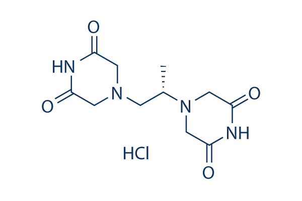
Struttura chimica
Peso molecolare: 304.73
Controllo Qualità
- Citato su Nature Medicine per la sua qualità superiore
- COA
- Scheda dati
- SDS
- Citato su Nature Medicine per la sua qualità superiore
- COA
- NMR
- Scheda dati
- SDS
- Citato su Nature Medicine per la sua qualità superiore
- COA
- NMR
- Scheda dati
- SDS
| Target correlati | HDAC PARP ATM/ATR DNA-PK WRN DNA/RNA Synthesis PPAR Sirtuin Casein Kinase eIF |
|---|---|
| Altro Topoisomerase Inibitori | Camptothecin (CPT) Betulinic acid Beta-Lapachone (S)-10-Hydroxycamptothecin Amonafide Voreloxin (SNS-595) hydrochloride Ellagic acid Cu(II)-Elesclomol Hydroxy Camptothecine Rubitecan |
Informazioni chimiche, conservazione e stabilità
| Peso molecolare | 304.73 | Formula | C11H16N4O4.HCl |
Conservazione (Dalla data di ricezione) | 3 years -20°C powder (seal) |
|---|---|---|---|---|---|
| N. CAS | 149003-01-0 | Scarica SDF | Conservazione delle soluzioni stock |
|
|
| Sinonimi | ADR-529 HCl, ICRF-187 HCl | Smiles | CC(CN1CC(=O)NC(=O)C1)N2CC(=O)NC(=O)C2.Cl | ||
Solubilità
|
In vitro |
DMSO
: 61 mg/mL
(200.17 mM)
Water : 61 mg/mL Ethanol : Insoluble |
Calcolatore di Molarità
|
In vivo |
|||||
Calcolatore di formulazione in vivo (Soluzione chiara)
Passo 1: Inserire le informazioni di seguito (Consigliato: Un animale aggiuntivo per tenere conto della perdita durante lesperimento)
Passo 2: Inserire la formulazione in vivo (Questo è solo il calcolatore, non la formulazione. Contattateci prima se non cè una formulazione in vivo nella sezione Solubilità.)
Risultati del calcolo:
Concentrazione di lavoro: mg/ml;
Metodo per preparare il liquido master di DMSO: mg farmaco predissolto in μL DMSO ( Concentrazione del liquido master mg/mL, Vi preghiamo di contattarci prima se la concentrazione supera la solubilità del DMSO del lotto del farmaco. )
Metodo per preparare la formulazione in vivo: Prendere μL DMSO liquido master, quindi aggiungereμL PEG300, mescolare e chiarire, quindi aggiungereμL Tween 80, mescolare e chiarire, quindi aggiungere μL ddH2O, mescolare e chiarire.
Metodo per preparare la formulazione in vivo: Prendere μL DMSO liquido master, quindi aggiungere μL Olio di mais, mescolare e chiarire.
Nota: 1. Si prega di assicurarsi che il liquido sia limpido prima di aggiungere il solvente successivo.
2. Assicurarsi di aggiungere il/i solvente/i in ordine. È necessario assicurarsi che la soluzione ottenuta, nellaggiunta precedente, sia una soluzione limpida prima di procedere allaggiunta del solvente successivo. Metodi fisici come il vortex, gli ultrasuoni o il bagno dacqua calda possono essere utilizzati per facilitare la dissoluzione.
Meccanismo dazione
| Targets/IC50/Ki |
Topo II
(Cell-free assay) |
|---|---|
| In vitro |
Il Dexrazoxane (10 mM), clinicamente noto per limitare la tossicità cardiaca da antracicline, previene l'apoptosi dei miociti indotta dalla daunorubicina, ma non la necrosi indotta da concentrazioni più elevate di antracicline nei miociti cardiaci di ratto. Il Dexrazoxane esercita presumibilmente i suoi effetti cardioprotettivi legando il ferro libero o debolmente legato, o il ferro complessato con doxorubicina, prevenendo o riducendo così la produzione di radicali ossigeno sito-specifici che danneggiano i componenti cellulari. Il Dexrazoxane abolisce specificamente il segnale di danno al DNA gamma-H2AX indotto dalla doxorubicina, ma non dalla camptotecina o dal perossido di idrogeno, nei cardiomiociti H9C2. Il Dexrazoxane induce anche una rapida degradazione di Top2beta, che è parallela alla riduzione del danno al DNA indotto dalla doxorubicina. Il Dexrazoxane antagonizza il danno al DNA indotto dalla doxorubicina attraverso la sua interferenza con Top2beta, il che potrebbe implicare Top2beta nella cardiotossicità da doxorubicina. Il Dexrazoxane viene idrolizzato nella sua forma attiva intracellularmente e lega il ferro per prevenire la formazione di radicali superidrossido, prevenendo così la distruzione mitocondriale. |
| In vivo |
Il Dexrazoxane combinato con doxorubicina, daunorubicina o idarubicina riduce le lesioni tissutali nei topi B6D2F1 (espresse come area sotto la curva della dimensione della ferita moltiplicata per la durata) del 96%, 70% e 87% rispettivamente. Il Dexrazoxane combinato con doxorubicina, daunorubicina o idarubicina si traduce in una riduzione statisticamente significativa della frazione di topi con ferite e della durata delle ferite. |
Riferimenti |
|
Applicazioni
| Metodi | Biomarcatori | Immagini | PMID |
|---|---|---|---|
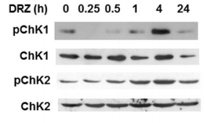
| Western blot | p-Chk1 / Chk1 / p-Chk2 / ChK2 ATF3 / TOP2A TOPOIIα / TOPOIIβ / TOPO I / PABP2 |
|
25521189 |
Supporto tecnico
Istruzioni per la manipolazione
Tel: +1-832-582-8158 Ext:3
Per qualsiasi altra domanda, si prega di lasciare un messaggio.

I prodotti sono solo per uso di ricerca. Non per uso umano. Non vendiamo a pazienti.
©Copyright 2013 Selleck Chemicals. Tutti i diritti riservati.