solo per uso di ricerca
BAY 11-7082 (BAY 11-7821) Inibitore di NF-κB
N. Cat.S2913
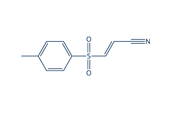
Struttura chimica
Peso molecolare: 207.25
Controllo Qualità
| Target correlati | HDAC Antioxidant ROS IκB/IKK Nrf2 AP-1 MALT NOD |
|---|---|
| Altro NF-κB Inibitori | DCZ0415 Omaveloxolone (RTA-408) JSH-23 QNZ (EVP4593) Caffeic Acid Phenethyl Ester SC75741 DHA (Dihydroartemisinin) Withaferin A (WFA) Andrographolide Evodiamine |
Coltura cellulare, trattamento e concentrazione di lavoro
| Linee cellulari | Tipo di saggio | Concentrazione | Tempo di incubazione | Formulazione | Descrizione dellattività | PMID |
|---|---|---|---|---|---|---|
| HeLa | Function Assay | 10 μM | 1.5 h | abolishes BPA induced up regulation of FN and MMP-9 | 25797437 | |
| SiHa | Function Assay | 10 μM | 1.5 h | abolishes BPA induced up regulation of FN and MMP-9 | 25797437 | |
| ARPE-19 | Function Assay | 1 μM | 0.5 h | suppresses TG-induced IL-8 promoter activation | 25593029 | |
| HCT116 | Function Assay | 5 μM | 2 h | DMSO | attenuates silymarin-induced downregulation of cyclin D1 | 25479723 |
| HMECs | Function Assay | 5 μM | 2 h | abolishes TNF-α-induced VCAM-1 expression | 25193116 | |
| A549 | Function Assay | 10 µM | 12 h | suppresses Dvl-3 induced activation of p65 | 25156800 | |
| RAW 264.7 | Function Assay | 5 μM | 1 h | inhibits TNF-α and IL-12 p40 production | 25019567 | |
| macrophages | Function Assay | 5 µM | 3 h | partially blocks YPFS-induced expression of iNOS and COX-2 | 24967898 | |
| HUVECs | Function Assay | 3-30 μM | 1 h | reduces the expression of miR-146a in a dose-dependent manner | 24863965 | |
| HeLa | Function Assay | 5 μM | 24 h | DMSO | reduces the activity of TNF-α promoter | 24657783 |
| A549 | Function Assay | 10 μM | 1 h | inhibits the increase of phospho-IκBα in PA103-infected cultures | 24612488 | |
| HUVEC | Function Assay | 20 µM | 0.5 h | DMSO | prevents the induction of EAM expression | 24551209 |
| A549RT-eto | Apoptosis Assay | 10 μM | 24 h | DMSO | accelerates FERO-mediated apoptosis | 24535083 |
| THP-1 | Function Assay | 0.1/1 μM | 0.5 h | abrogates TNF-α secretion as well as the increased secretion of IL-6 and IL-1β | 24378536 | |
| SKCXCR2 | Growth Inhibition Assay | 2 µM | 48 h | decreases cell proliferation significantly | 24376747 | |
| SKCXCR2 | Function Assay | 2 µM | 48 h | blocks the CXCL1-induced cell invasion | 24376747 | |
| OVCXCR2 | Function Assay | 2 µM | 48 h | blocks the CXCL1-induced cell invasion | 24376747 | |
| DSCs | Function Assay | 2.5 μM | 0.5 h | reverses the enhancement of CCL2/CCR2 expression of DSCs induced by IL-33 | 24344240 | |
| WPs | Function Assay | 25 μM | 5 min | suppresses ATP and vWF secretion | 24331207 | |
| A549RT-eto | Apoptosis Assay | 10 μM | 24 h | accelerates F14 extract-mediated apoptosis combined treatment with F14 | 24220725 | |
| A549RT-eto | Function Assay | 10 μM | 24 h | decreases the expression levels of NF-κB and P-gp | 24220725 | |
| FaDu | Function Assay | 2 h | inhibits p65 expression and blocks TNFα-induced TWIST expression | 24220622 | ||
| IVD | Function Assay | 10 μM | 3 d | reverses TNF-α–mediated suppression of the disc matrix macromolecules aggrecan and collagen II | 24176808 | |
| IVD | Function Assay | 10 μM | 3 d | abrogates TNF-α–induced up-regulation of ADAMTS-4 and ADAMTS-5 | 24176808 | |
| iNKT | Function Assay | 10/100 μM | 0.5 h | inhibits the induction of A2AR mRNA and other factor | 24124453 | |
| PC-3 | Function Assay | 2.5/5/10 μM | 0.5 h | blocks IGF-II-induced STS mRNA expression | 24055520 | |
| THP-1 | Function Assay | 10 μM | 1 h | abolishes the effect of rHSP27 on SR-A mRNA | 23939398 | |
| A549 | Function Assay | 1 μM | 48 h | enhances the up-regulation of IκB and subsequent decrease in Bax expression induced by combined stimulation | 23900080 | |
| A549 | Apoptosis Assay | 1 μM | 48 h | reduces the cell death induced by combined stimulation | 23900080 | |
| NCI-N87 | Growth Inhibition Assay | 10/20/30 μM | 6/24 h | suppresses cell viability significantly | 23846545 | |
| AGS | Growth Inhibition Assay | 10/20/30 μM | 6/24 h | suppresses cell viability significantly | 23846545 | |
| MGC80-3 | Growth Inhibition Assay | 10/20/30 μM | 6/24 h | suppresses cell viability significantly | 23846545 | |
| HGC-27 | Function Assay | 7.5/15/30 μM | 6 h | induces the dephosphorylation and up-regulation of IκBα | 23846545 | |
| MGC80-3 | Function Assay | 7.5/15/30 μM | 6 h | induces the dephosphorylation and up-regulation of IκBα | 23846545 | |
| HGC-27 | Apoptosis Assay | 7.5/15/30 μM | 6 h | induces apoptosis in a time- and dose-dependent manner | 23846545 | |
| HBE | Function Assay | 10μM | 3h | abolishes the increases of IL-6 expression induced by CSE | 23824089 | |
| HepG2 | Function Assay | 0.3/1/3 μM | 48 h | reduces IL6-induced PON1 expression | 23791833 | |
| THP-1 | Function Assay | 5 µM | 1 h | DMSO | inhibits MTB-induced NFκB activation | 23634218 |
| THP-1 | Growth Inhibition Assay | 5 µM | 4/8 d | DMSO | reduces the viability of intracellular MTB | 23634218 |
| MDM | Growth Inhibition Assay | 5 µM | 4/8 d | DMSO | reduces the viability of intracellular MTB | 23634218 |
| AM | Growth Inhibition Assay | 5 µM | 4/8 d | DMSO | reduces the viability of intracellular MTB | 23634218 |
| RAW 264 | Function Assay | 0.2-5 µM | 30/60/90 min | inhibits the phosphatase activity of PTP1B | 23578302 | |
| HUVEC | Function Assay | 10 μM | 0.5 h | DMSO | counteractes the loss of Tie2 mRNA | 23563632 |
| HT29 | Function Assay | 10/30/100 μM | 1 h | inhibites both TWEAK-induced p100 processing | 23527154 | |
| HT29 | Function Assay | 10/30/100 μM | 1 h | inhibits TNF-induced phosphorylation and degradation of IκBα | 23527154 | |
| MM.1S | Apoptosis Assay | 30 µM | 3 h | induces MM cell death involves necrosis | 23527154 | |
| KMS-12-BM | Apoptosis Assay | 30 µM | 3 h | induces MM cell death involves necrosis | 23527154 | |
| BAFs | Function Assay | 0.5/1 μM | 24 h | inhibits TNFα/DEX induced CYP19A1 transcripts | 23485457 | |
| SP6.5 | Function Assay | 5 μM | 2 h | decreases translocation of p65 in the nucleus | 23443086 | |
| VUP | Function Assay | 5 μM | 2 h | decreases translocation of p65 in the nucleus | 23443086 | |
| OCM1 | Function Assay | 5 μM | 2 h | decreases translocation of p65 in the nucleus | 23443086 | |
| OM431 | Function Assay | 5 μM | 2 h | decreases translocation of p65 in the nucleus | 23443086 | |
| SP6.5 | Growth Inhibition Assay | 2.5-20 μM | 24 h | IC50=5 μM, exhibits strong anti-proliferative effects in a dose-dependent manner | 23443086 | |
| VUP | Growth Inhibition Assay | 2.5-20 μM | 24 h | IC50=5 μM, exhibits strong anti-proliferative effects in a dose-dependent manner | 23443086 | |
| OCM1 | Growth Inhibition Assay | 2.5-20 μM | 24 h | IC50=5 μM, exhibits strong anti-proliferative effects in a dose-dependent manner | 23443086 | |
| OM431 | Growth Inhibition Assay | 2.5-20 μM | 24 h | IC50=5 μM, exhibits strong anti-proliferative effects in a dose-dependent manner | 23443086 | |
| SP6.5 | Apoptosis Assay | 5 μM | 24 h | induces apoptosis | 23443086 | |
| VUP | Apoptosis Assay | 5 μM | 24 h | induces apoptosis | 23443086 | |
| OCM1 | Apoptosis Assay | 5 μM | 24 h | induces apoptosis | 23443086 | |
| OM431 | Apoptosis Assay | 5 μM | 24 h | induces apoptosis | 23443086 | |
| SP6.5 | Function Assay | 5 μM | 12 h | reduces the migration | 23443086 | |
| VUP | Function Assay | 5 μM | 12 h | reduces the migration | 23443086 | |
| OCM1 | Function Assay | 5 μM | 12 h | reduces the migration | 23443086 | |
| OM431 | Function Assay | 5 μM | 12 h | reduces the migration | 23443086 | |
| HBL-1 | Growth Inhibition Assay | 3 μM | 24/48/72 h | DMSO | slows cell growth modestly | 23441730 |
| RAW 264.7 | Function Assay | 2-15 μM | 1 h | DMSO | suppresses the activation of IKK family members | 23441730 |
| IL-1R | Function Assay | 2-15 μM | 1 h | DMSO | suppresses the activation of IKK family members | 23441730 |
| RAW 264.7 | Function Assay | 15 μM | 1 h | DMSO | suppresses the activation of and JNK | 23441730 |
| IL-1R | Function Assay | 15 μM | 1 h | DMSO | suppresses the activation of and JNK | 23441730 |
| U2OS | Function Assay | 15 μM | 1 h | DMSO | prevents the LPS- or IL-1-stimulated formation of K63-pUb chains | 23441730 |
| MT‐1 | Function Assay | 8 µm | 3 h | decreases the levels of p‐STAT3 and p‐4EBP1 | 23278479 | |
| MT‐2 | Function Assay | 8 µm | 3 h | decreases the levels of p‐STAT3 and p‐4EBP1 | 23278479 | |
| MT‐1 | Function Assay | 8 µm | 3 h | decreases the levels of the p65 subunit of NF‐κB | 23278479 | |
| MT‐2 | Function Assay | 8 µm | 3 h | decreases the levels of the p65 subunit of NF‐κB | 23278479 | |
| MCF-7 | Function Assay | 2.5-15 μM | 0.5 h | DMSO | causes the gradual loss of cell adhesion | 23093227 |
| HaCaT | Function Assay | 5.0 μM | 1 h | attenuates the TCOH-induced production of IL-6 | 23041168 | |
| A549 | Function Assay | 1 h | inhibits LTA-induced SP-A mRNA production significantly | 23031213 | ||
| OA chondrocytes | Function Assay | 10 μM | 1 h | blocks the AGE-BSA-induced gene/protein expression of GRP78 or COX-2 (p<0.05) | 22982228 | |
| RAW264.7 | Function Assay | 15 μM | 15-120 min | blocks the production of NO, PGE2, and TNF-α | 22745523 | |
| RAW264.7 | Growth Inhibition Assay | 5-30 μM | 24 h | inhibits cell growth in a dose-dependent manner | 22745523 | |
| HBL6 | Apoptosis Assay | 0.5/5/25 μM | 6/24 h | decreases cell viability and leeads to apoptosis in a dose-dependent manner | 22074820 | |
| HT29 | Function Assay | 1-10 μM | 10 h | increases HO-1 mRNA and protein expression | 21620964 | |
| Ca9–22 | Apoptosis Assay | 10 μM | 1 h | completely inhibits ALA-PDT-induced apoptosis | 21138480 | |
| Ca9–22 | Function Assay | 10 μM | 1 h | completely abrogates the ALA-PDT-induced JNK activation | 21138480 | |
| A-549 | Growth Inhibition Assay | 10 μM | 24/48 h | inhibits cell growth in a time-dependent manner | 20866043 | |
| AP | Function Assay | 5/10 μM | 48 h | downregulates the BAD protein level a dose-dependent manner | 20596645 | |
| AQ1 | Function Assay | 5/10 μM | 48 h | downregulates the BAD protein level a dose-dependent manner | 20596645 | |
| AP | Function Assay | 20 μM | 4/8 h | downregulates the BAD protein level a time-dependent manner | 20596645 | |
| AQ1 | Function Assay | 20 μM | 4/8 h | downregulates the BAD protein level a time-dependent manner | 20596645 | |
| THP-1 | Function Assay | 5 μM | 0.5 h | attenuates the LPS-induced p-IκBα protein by 72% | 20309718 | |
| K562 | Growth Inhibition Assay | 2-30 μM | 24 h | IC50=8 μM,inhibits cell growth in a dose-dependent manner | 19646807 | |
| Jurket | Growth Inhibition Assay | 2-30 μM | 24 h | IC50=7.1 μM, inhibits cell growth in a dose-dependent manner | 19646807 | |
| U937 | Growth Inhibition Assay | 2-30 μM | 24 h | IC50=10.5 μM, inhibits cell growth in a dose-dependent manner | 19646807 | |
| PBMC | Growth Inhibition Assay | 2-30 μM | 24 h | IC50=40.2 μM, inhibits cell growth in a dose-dependent manner | 19646807 | |
| K562 | Apoptosis Assay | 2-20 μM | 24 h | induces a dose-dependent apoptosis | 19646807 | |
| THP1 | Cytotoxicity assay | 72 hrs | Cytotoxicity against human THP1 cells assessed as reduction in cell viability after 72 hrs by MTT assay, TC50 = 1.5 μM. | 28410442 | ||
| RAW264.7 | Function assay | 6 hrs | Inhibition of LPS-induced NF-kappaB activation in mouse RAW264.7 cells treated 30 mins before LPS challenge measured after 6 hrs by luciferase reporter gene assay, IC50 = 1.72 μM. | 24315191 | ||
| HEK293 | Function assay | 6 hrs | Inhibition of TNF-alpha-induced NF-kappaB activity in HEK293 cells after 6 hrs by luciferase reporter gene assay, IC50 = 2 μM. | 24533857 | ||
| HEK293 | Function assay | 6 hrs | Inhibition of TNF-alpha-induced NF-kappaB activity in HEK293 cells after 6 hrs by luciferase reporter gene assay, IC50 = 2 μM. | 24992702 | ||
| HEK293 | Function assay | 6 hrs | Inhibition of TNFalpha-induced NF-kappaB activity (unknown origin) transfected in HEK293 cells after 6 hrs by luciferase reporter gene assay, IC50 = 2 μM. | 26343828 | ||
| HEK293 | Function assay | 6 hrs | Inhibition of TNFalpha-induced NF-kappaB activity expressed in human HEK 293 cells after 6 hrs by luciferase reporter gene assay, IC50 = 2 μM. | 22850207 | ||
| HEK293 | Function assay | 6 hrs | Inhibition of TNFalpha-induced human NFkappaB activity in HEK293 cells incubated for 6 hrs followed by compound wash out measured after 5 mins by by luciferase assay, IC50 = 2.01 μM. | 22712432 | ||
| HEK293 | Cytotoxicity assay | Cytotoxicity against HEK293 cells, IC50 = 3.8 μM. | 24533857 | |||
| HEK293 | Function assay | 6 hrs | Inhibition of TNFalpha-induced NFkappaB (unknown origin) activation expressed in HEK293 cells after 6 hrs by luciferase reporter gene assay, IC50 = 5 μM. | 23316950 | ||
| HEK293 | Function assay | Effect on Cdc2 expressed in HEK293 cells assessed as effect on Cdc2:Cdc25C interaction complexes in presence of camptothecin by EYFP and/or YFP Venus fragment based reporter gene assay | 16680159 | |||
| HEK293 | Function assay | 20 uM | 24 hrs | Inhibition of TNF-alpha stimulated NFkappaB transactivation in HEK293 cells at 20 uM measured after 24 hrs by dual luciferase reporter gene assay | 27736063 | |
| RAW264.7 | Function assay | 20 uM | 1 hr | Inhibition of LPS-induced NFkB activation in mouse RAW264.7 cells assessed as reduction in nuclear translocation of p65 at 20 uM preincubated for 1 hr followed by LPS stimulation measured after 3 hrs by Western blot method | 28667873 | |
| RAW264.7 | Function assay | 20 uM | 6 hrs | Inhibition of LPS-induced NF-kappaB activation in mouse RAW264.7 cells at 20 uM treated 30 mins before LPS challenge measured after 6 hrs by luciferase reporter gene assay | 24315191 | |
| RAW264.7 | Antinflammatory assay | 20 uM | 18 hrs | Antinflammatory activity in mouse RAW264.7 cells assessed as inhibition of LPS-induced nitric oxide production at 20 uM treated 30 mins before LPS challenge measured after 18 hrs by Griess assay | 24315191 | |
| THP1 | Antinflammatory assay | 5 uM | 24 hrs | Antiinflammatory activity in human THP1 cells assessed as inhibition of TPA/ionomycin-induced extracellular IL-1beta level at 5 uM incubated 1 hr prior to TPA/ionomycin challenge measured after 24 hrs by ELISA | 24400858 | |
| THP1 | Antinflammatory assay | 5 uM | 24 hrs | Antiinflammatory activity in human THP1 cells assessed as inhibition of TPA/ionomycin-induced extracellular TNF-alpha production at 5 uM incubated 1 hr prior to TPA/ionomycin challenge measured after 24 hrs by ELISA | 24400858 | |
| THP1 | Antinflammatory assay | 5 uM | 24 hrs | Antiinflammatory activity in human THP1 cells assessed as inhibition of TPA/ionomycin-induced intracellular proIL-1beta level at 5 uM incubated 1 hr prior to TPA/ionomycin challenge measured after 24 hrs by ELISA | 24400858 | |
| THP1 | Antinflammatory assay | 5 uM | 24 hrs | Antiinflammatory activity in human THP1 cells assessed as inhibition of TPA/ionomycin-induced intracellular IL-1beta level at 5 uM incubated 1 hr prior to TPA/ionomycin challenge measured after 24 hrs by ELISA | 24400858 | |
| RAW264.7 | Function assay | 0.3 ug/ml | 12 hrs | Inhibition of LPS-induced NF-kB p65 phosphorylation in mouse RAW264.7 cells at 0.3 ug/ml preincubated for 12 hrs followed by LPS stimulation for 3 hrs by Western blot method | 28284806 | |
| RAW264.7 | Function assay | 0.3 ug/ml | 12 hrs | Inhibition of LPS-induced NF-kB p65 activation in mouse RAW264.7 cells at 0.3 ug/ml preincubated for 12 hrs followed by LPS stimulation for 3 hrs by DAPI staining based inverted fluorescence microscopic method | 28284806 | |
| RAW264.7 | Function assay | 0.3 ug/ml | 12 hrs | Inhibition of NF-kB p65 in mouse RAW264.7 cells assessed as reduction in LPS-induced iNOS expression at 0.3 ug/ml preincubated for 12 hrs followed by LPS stimulation for 3 hrs by Western blot method | 28284806 | |
| RAW264.7 | Function assay | 0.3 ug/ml | 12 hrs | Inhibition of NF-kB p65 in mouse RAW264.7 cells assessed as reduction in LPS-induced COX2 expression at 0.3 ug/ml preincubated for 12 hrs followed by LPS stimulation for 3 hrs by Western blot method | 28284806 | |
| HEK293 | Function assay | 20 uM | 24 hrs | Inhibition of TNFalpha-induced NFkappaB activation in HEK293 cells at 20 uM after 24 hrs by dual luciferase reporter gene assay | 28873303 | |
| RAW264.7 | Function assay | 20 uM | 2 hrs | Inhibition of NFkappaB nuclear translocation in LPS-stimulated mouse RAW264.7 cells at 20 uM pretreated for 2 hrs followed by LPS-induction by DAPI-staining based immunofluorescence microscopic method | 29759725 | |
| BGC823 | Function assay | 5 uM | 12 hrs | Inhibition of colony formation in human BGC823 cells at 5 uM treated for 12 hrs followed by incubation in drug free medium for 14 days by crystal violet staining based assay | 28881286 | |
| SGC7901 | Function assay | 5 uM | 12 hrs | Inhibition of colony formation in human SGC7901 cells at 5 uM treated for 12 hrs followed by incubation in drug free medium for 14 days by crystal violet staining based assay | 28881286 | |
| RAW264.7 | Function assay | 10 uM | 2 hrs | Inhibition of LPS-induced IL-6 mRNA expression in mouse RAW264.7 cells at 10 uM pre-incubated for 2 hrs before LPS stimulation for 24 hrs by qRT-PCR method | 27038497 | |
| RAW264.7 | Function assay | 10 uM | 2 hrs | Inhibition of LPS-induced IL-1beta mRNA expression in mouse RAW264.7 cells at 10 uM pre-incubated for 2 hrs before LPS stimulation for 24 hrs by qRT-PCR method | 27038497 | |
| RAW264.7 | Function assay | 10 uM | 2 hrs | Inhibition of LPS-induced iNOS mRNA expression in mouse RAW264.7 cells at 10 uM pre-incubated for 2 hrs before LPS stimulation for 24 hrs by qRT-PCR method | 27038497 | |
| Clicca per visualizzare più dati sperimentali sulle linee cellulari | ||||||
Informazioni chimiche, conservazione e stabilità
| Peso molecolare | 207.25 | Formula | C10H9NO2S |
Conservazione (Dalla data di ricezione) | |
|---|---|---|---|---|---|
| N. CAS | 19542-67-7 | Scarica SDF | Conservazione delle soluzioni stock |
|
|
| Sinonimi | BAY 11-7821 | Smiles | CC1=CC=C(C=C1)S(=O)(=O)C=CC#N | ||
Solubilità
|
In vitro |
DMSO
: 41 mg/mL
(197.82 mM)
Ethanol : 10 mg/mL Water : Insoluble |
Calcolatore di Molarità
|
In vivo |
|||||
Calcolatore di formulazione in vivo (Soluzione chiara)
Passo 1: Inserire le informazioni di seguito (Consigliato: Un animale aggiuntivo per tenere conto della perdita durante lesperimento)
Passo 2: Inserire la formulazione in vivo (Questo è solo il calcolatore, non la formulazione. Contattateci prima se non cè una formulazione in vivo nella sezione Solubilità.)
Risultati del calcolo:
Concentrazione di lavoro: mg/ml;
Metodo per preparare il liquido master di DMSO: mg farmaco predissolto in μL DMSO ( Concentrazione del liquido master mg/mL, Vi preghiamo di contattarci prima se la concentrazione supera la solubilità del DMSO del lotto del farmaco. )
Metodo per preparare la formulazione in vivo: Prendere μL DMSO liquido master, quindi aggiungereμL PEG300, mescolare e chiarire, quindi aggiungereμL Tween 80, mescolare e chiarire, quindi aggiungere μL ddH2O, mescolare e chiarire.
Metodo per preparare la formulazione in vivo: Prendere μL DMSO liquido master, quindi aggiungere μL Olio di mais, mescolare e chiarire.
Nota: 1. Si prega di assicurarsi che il liquido sia limpido prima di aggiungere il solvente successivo.
2. Assicurarsi di aggiungere il/i solvente/i in ordine. È necessario assicurarsi che la soluzione ottenuta, nellaggiunta precedente, sia una soluzione limpida prima di procedere allaggiunta del solvente successivo. Metodi fisici come il vortex, gli ultrasuoni o il bagno dacqua calda possono essere utilizzati per facilitare la dissoluzione.
Meccanismo dazione
| Targets/IC50/Ki |
E2-conjugating enzymes
(Cell-free assay) USP7
(Cell-free assay) 0.19 μM
USP21
(Cell-free assay) 0.96 μM
USP6
(Cell-free assay) 1.7 μM
IκBα phosphorylation
(Tumor cells) 10 μM
|
|---|---|
| In vitro |
BAY 11-7082 abroga completamente e specificamente il legame del DNA di NF-κB, downregolando la citochina inducibile da NF-κB IL-6 e inducendo l'apoptosi. Questo composto (< 8 μM) è in grado di inibire efficacemente l'attività luciferasica di NFκB sia basale che stimolata da TNFα in modo dose-dipendente. Esso (8 μM) inibisce fortemente il tasso di proliferazione nelle cellule NCI-H1703. Questo composto (5 μM) riduce rapidamente ed efficacemente il legame del DNA di NF-kappaB nelle linee cellulari T infettate da HTLV-I e down-regola l'espressione del gene antiapoptotico, Bcl-x(L), mentre ha scarso effetto sul legame del DNA di un altro fattore di trascrizione, AP-1. Questa apoptosi indotta chimicamente delle cellule ATL primarie è più evidente di quella delle normali cellule mononucleate del sangue periferico, e l'apoptosi di queste cellule è anche associata alla down-regolazione dell'attività di NF-kappaB. Esso (5 μM) induce selettivamente l'apoptosi delle linee cellulari T infettate da HTLV-I associata alla down-regolazione dell'espressione di ciclina D1, ciclina D2 e Bcl-xL. Questo composto (100 μM) previene la traslocazione nucleare di p65 elicitata da NMDA e l'aumento indotto da NMDA del legame di NF-κB in sezioni di ippocampo di topo. Previene la tossicità da NMDA che si verifica nella regione CA1 di sezioni di ippocampo con una neuroprotezione del 40% a 20 μM e del 70% a 100 μM. Questa sostanza chimica a tutte le concentrazioni testate inibisce significativamente l'attività di legame del DNA di NF-κB p65 nel tessuto adiposo, mentre nel muscolo scheletrico, a 50 μM e 100 μM, inibisce significativamente l'attività di legame del DNA di NF-κB p65. Essa (100 μM) riduce la proteina IKK-β nel tessuto adiposo umano e nel muscolo scheletrico. Questo composto (100 μM) diminuisce significativamente il rilascio di TNF-α dal tessuto adiposo, mentre il rilascio di IL-6 e IL-8 è significativamente inibito a tutte le concentrazioni di questa sostanza chimica testate. Esso (50 μM) diminuisce significativamente il rilascio di TNF-α, IL-6 e IL-8 nel muscolo scheletrico. Questo composto è anche risultato inattivare gli enzimi di coniugazione E2 Ubc (ubiquitina coniugante) 13 e UbcH7 e la ligasi E3 LUBAC (linear ubiquitin assembly complex), e quindi induce la morte di linfomi a cellule B e leucemie a cellule T. |
| In vivo |
BAY 11-7082, un inibitore di NF-κB, induce apoptosi e arresto della fase S nelle cellule di cancro gastrico. |
Riferimenti |
|
Applicazioni
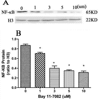
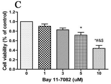
| Metodi | Biomarcatori | Immagini | PMID |
|---|---|---|---|
| Western blot | NF-κB p-IKKβ/ IκBα p-IRAK4 / IRAK4 NF-κB p-IKKβ/ IκBα p-IRAK4 / IRAK4 |
|
31332209 |
| Growth inhibition assay | Cell viability Cell viability |
|
31332209 |
Supporto tecnico
Istruzioni per la manipolazione
Tel: +1-832-582-8158 Ext:3
Per qualsiasi altra domanda, si prega di lasciare un messaggio.
I prodotti sono solo per uso di ricerca. Non per uso umano. Non vendiamo a pazienti.
©Copyright 2013 Selleck Chemicals. Tutti i diritti riservati.