
solo per uso di ricerca
Vadimezan (DMXAA) Agente Disruptore Vascolare
N. Cat.S1537
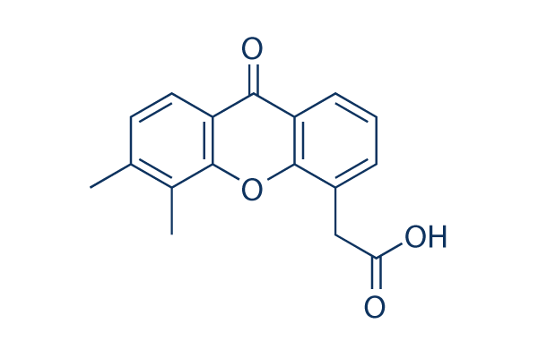
Struttura chimica
Peso molecolare: 282.29
Controllo Qualità
Coltura cellulare, trattamento e concentrazione di lavoro
| Linee cellulari | Tipo di saggio | Concentrazione | Tempo di incubazione | Formulazione | Descrizione dellattività | PMID |
|---|---|---|---|---|---|---|
| human BJ cells | Cytotoxic assay | 24 h | Cytotoxicity against human BJ cells after 24 hrs by MTT assay, CC50=48.9 μM | 24518295 | ||
| HECPP cells | Function assay | 10 ug/mL | Activation of NF-kappaB in HECPP cells at 10 ug/mL | 17616114 | ||
| MCF7 | Antiproliferative assay | 24 hrs | Antiproliferative activity against human MCF7 cells co-treated with pyranoxanthone at 1:1 molar ratio after 24 hrs by MTT assay, IC50 = 11.89 μM. | 29129511 | ||
| MDA-MB-231 | Antiproliferative assay | 24 hrs | Antiproliferative activity against human MDA-MB-231 cells co-treated with pyranoxanthone at 1:1 molar ratio after 24 hrs by MTT assay, IC50 = 12.12 μM. | 29129511 | ||
| K562 | Antiproliferative assay | 24 hrs | Antiproliferative activity against human K562 cells co-treated with pyranoxanthone at 1:1 molar ratio after 24 hrs by MTT assay, IC50 = 19.14 μM. | 29129511 | ||
| HepG2 | Antiproliferative assay | 24 hrs | Antiproliferative activity against human HepG2 cells co-treated with pyranoxanthone at 1:1 molar ratio after 24 hrs by MTT assay, IC50 = 21.25 μM. | 29129511 | ||
| COLO320 | Antiproliferative assay | 48 hrs | Antiproliferative activity against human COLO320 cells after 48 hrs by CCK8 assay, IC50 = 39.5 μM. | 28376372 | ||
| MDA-MB-231 | Antiproliferative assay | 48 hrs | Antiproliferative activity against human MDA-MB-231 cells after 48 hrs by CCK8 assay, IC50 = 48.4 μM. | 28376372 | ||
| MDA-MB-231 | Growth inhibition assay | 24 hrs | Growth inhibition of human MDA-MB-231 cells after 24 hrs by MTT assay, IC50 = 48.42 μM. | 29609121 | ||
| MDA-MB-231 | Antiproliferative assay | 24 hrs | Antiproliferative activity against human MDA-MB-231 cells after 24 hrs by MTT assay, IC50 = 48.44 μM. | 29129511 | ||
| A673 | qHTS assay | qHTS of pediatric cancer cell lines to identify multiple opportunities for drug repurposing: Primary screen for A673 cells | 29435139 | |||
| DAOY | qHTS assay | qHTS of pediatric cancer cell lines to identify multiple opportunities for drug repurposing: Primary screen for DAOY cells | 29435139 | |||
| RD | qHTS assay | qHTS of pediatric cancer cell lines to identify multiple opportunities for drug repurposing: Primary screen for RD cells | 29435139 | |||
| SK-N-SH | qHTS assay | qHTS of pediatric cancer cell lines to identify multiple opportunities for drug repurposing: Primary screen for SK-N-SH cells | 29435139 | |||
| MG 63 (6-TG R) | qHTS assay | qHTS of pediatric cancer cell lines to identify multiple opportunities for drug repurposing: Primary screen for MG 63 (6-TG R) cells | 29435139 | |||
| NB1643 | qHTS assay | qHTS of pediatric cancer cell lines to identify multiple opportunities for drug repurposing: Primary screen for NB1643 cells | 29435139 | |||
| Rh41 | qHTS assay | qHTS of pediatric cancer cell lines to identify multiple opportunities for drug repurposing: Primary screen for Rh41 cells | 29435139 | |||
| SK-N-MC | qHTS assay | qHTS of pediatric cancer cell lines to identify multiple opportunities for drug repurposing: Primary screen for SK-N-MC cells | 29435139 | |||
| MDA-MB-231 | Apoptosis assay | 24 to 96 uM | 48 hrs | Induction of apoptosis in human MDA-MB-231 cells assessed as increase in cleaved caspase-3 expression at 24 to 96 uM after 48 hrs by Western blot analysis | 28376372 | |
| MDA-MB-231 | Apoptosis assay | 24 to 96 uM | 48 hrs | Induction of apoptosis in human MDA-MB-231 cells assessed as increase in cleaved PARP level at 24 to 96 uM after 48 hrs by Western blot analysis | 28376372 | |
| MDA-MB-231 | Function assay | 24 to 96 uM | 48 hrs | Decrease in caspase-3 level in human MDA-MB-231 cells at 24 to 96 uM after 48 hrs by Western blot analysis | 28376372 | |
| MDA-MB-231 | Function assay | 24 to 96 uM | 48 hrs | Increase in p53 level in human MDA-MB-231 cells at 24 to 96 uM after 48 hrs by Western blot analysis | 28376372 | |
| MDA-MB-231 | Function assay | 24 to 96 uM | 48 hrs | Decrease in caspase-9 level in human MDA-MB-231 cells at 24 to 96 uM after 48 hrs by Western blot analysis | 28376372 | |
| MDA-MB-231 | Function assay | 24 to 96 uM | 48 hrs | Decrease in MDM2 level in human MDA-MB-231 cells at 24 to 96 uM after 48 hrs by Western blot analysis | 28376372 | |
| HepG2 | Cell cycle arrest assay | 0.2 uM | 24 hrs | Cell cycle arrest in human HepG2 cells assessed as accumulation at S phase at 0.2 uM after 24 hrs by propidium iodide staining-based flow cytometric method relative to control | 29129511 | |
| HepG2 | Cell cycle arrest assay | 24 hrs | Cell cycle arrest in human HepG2 cells assessed as accumulation at S phase co-treated with pyranoxanthone at 1:1 molar ratio after 24 hrs by propidium iodide staining-based flow cytometric method | 29129511 | ||
| HepG2 | Apoptosis assay | 0.2 uM | 24 hrs | Induction of apoptosis in human HepG2 cells assessed as increase in cleaved caspase-3 levels at 0.2 uM after 24 hrs by Western blot method | 29129511 | |
| HepG2 | Apoptosis assay | 0.2 uM | 24 hrs | Induction of apoptosis in human HepG2 cells assessed as increase in cleaved caspase-9 levels at 0.2 uM after 24 hrs by Western blot method | 29129511 | |
| HepG2 | Apoptosis assay | 24 hrs | Induction of apoptosis in human HepG2 cells assessed as increase in cleaved PARP levels co-treated with pyranoxanthone at 1:1 molar ratio after 24 hrs by Western blot method | 29129511 | ||
| HepG2 | Apoptosis assay | 0.2 uM | 24 hrs | Induction of apoptosis in human HepG2 cells assessed as increase in cleaved PARP levels at 0.2 uM after 24 hrs by Western blot method | 29129511 | |
| HepG2 | Apoptosis assay | 24 hrs | Induction of apoptosis in human HepG2 cells assessed as increase in cleaved caspase-3 levels co-treated with pyranoxanthone at 1:1 molar ratio after 24 hrs by Western blot method | 29129511 | ||
| HepG2 | Apoptosis assay | 24 hrs | Induction of apoptosis in human HepG2 cells assessed as increase in cleaved caspase-9 levels co-treated with pyranoxanthone at 1:1 molar ratio after 24 hrs by Western blot method | 29129511 | ||
| HepG2 | Apoptosis assay | 24 hrs | Induction of apoptosis in human HepG2 cells assessed as downregulation of Bcl-xL expression co-treated with pyranoxanthone at 1:1 molar ratio after 24 hrs by Western blot method | 29129511 | ||
| HepG2 | Apoptosis assay | 24 hrs | Induction of apoptosis in human HepG2 cells assessed as upregulation of Bid expression co-treated with pyranoxanthone at 1:1 molar ratio after 24 hrs by Western blot method | 29129511 | ||
| Clicca per visualizzare più dati sperimentali sulle linee cellulari | ||||||
Informazioni chimiche, conservazione e stabilità
| Peso molecolare | 282.29 | Formula | C17H14O4 |
Conservazione (Dalla data di ricezione) | |
|---|---|---|---|---|---|
| N. CAS | 117570-53-3 | Scarica SDF | Conservazione delle soluzioni stock |
|
|
| Sinonimi | ASA404, NSC 640488 | Smiles | CC1=C(C2=C(C=C1)C(=O)C3=CC=CC(=C3O2)CC(=O)O)C | ||
Solubilità
|
In vitro |
DMSO
: 16 mg/mL
(56.67 mM)
Riscaldato con bagno dacqua a 50°C;
Sonicato;
7.5%Sodium bicarbonate : 10 mg/mL (Ultrasonic and heating for 5 minutes.) Water : Insoluble |
Calcolatore di Molarità
|
In vivo |
|||||
Calcolatore di formulazione in vivo (Soluzione chiara)
Passo 1: Inserire le informazioni di seguito (Consigliato: Un animale aggiuntivo per tenere conto della perdita durante lesperimento)
Passo 2: Inserire la formulazione in vivo (Questo è solo il calcolatore, non la formulazione. Contattateci prima se non cè una formulazione in vivo nella sezione Solubilità.)
Risultati del calcolo:
Concentrazione di lavoro: mg/ml;
Metodo per preparare il liquido master di DMSO: mg farmaco predissolto in μL DMSO ( Concentrazione del liquido master mg/mL, Vi preghiamo di contattarci prima se la concentrazione supera la solubilità del DMSO del lotto del farmaco. )
Metodo per preparare la formulazione in vivo: Prendere μL DMSO liquido master, quindi aggiungereμL PEG300, mescolare e chiarire, quindi aggiungereμL Tween 80, mescolare e chiarire, quindi aggiungere μL ddH2O, mescolare e chiarire.
Metodo per preparare la formulazione in vivo: Prendere μL DMSO liquido master, quindi aggiungere μL Olio di mais, mescolare e chiarire.
Nota: 1. Si prega di assicurarsi che il liquido sia limpido prima di aggiungere il solvente successivo.
2. Assicurarsi di aggiungere il/i solvente/i in ordine. È necessario assicurarsi che la soluzione ottenuta, nellaggiunta precedente, sia una soluzione limpida prima di procedere allaggiunta del solvente successivo. Metodi fisici come il vortex, gli ultrasuoni o il bagno dacqua calda possono essere utilizzati per facilitare la dissoluzione.
Meccanismo dazione
| Targets/IC50/Ki |
DT-diaphorase
(Cell-free assay) 20 μM(Ki)
DT-diaphorase
(Cell-free assay) 20 μM(Ki)
|
|---|---|
| In vitro |
Nelle cellule di carcinoma del colon umano DLD-1, Vadimezan (DMXAA) inibisce l'attività della DT-diaphorase senza effetti significativi sull'attività della citocromo b5 reduttasi e della citocromo P450 reduttasi. La combinazione di menadione e questo composto porta ad un aumento dell'attività antiproliferativa delle cellule DLD-1. Come agente Antiviral, inibisce la citotossicità indotta da VSV e la replicazione del virus influenzale nei macrofagi RAW 264.7. Uno studio recente mostra che DMXAA ha effetti inibitori non immuno-mediati contro diversi membri chinasici del VEGFR (recettore del fattore di crescita endoteliale vascolare), come la segnalazione di VEGFR2 nelle cellule endoteliali della vena ombelicale umana.
|
| Saggio chinasico |
Attività della DT-diaphorase e analisi cinetica dell'inibizione enzimatica
|
|
L'attività dell'enzima DT-diaphorase purificata viene saggiata misurando la riduzione del citocromo c a 550 nm su uno spettrofotometro Beckman DU 650. Ogni saggio contiene citocromo c (70 μM), NADH (concentrazioni variabili), DT-diaphorase purificata (0,032 μg) e menadione (concentrazioni variabili) in un volume finale di 1 mL di tampone Tris–HCl (50 mM, pH 7,4) contenente 0,14% di BSA. La reazione viene avviata con l'aggiunta di NADH. I tassi di riduzione vengono calcolati sulla parte iniziale della curva di reazione (30 secondi) e i risultati sono espressi in termini di μmol di citocromo c ridotto/min/mg di proteina utilizzando un coefficiente di estinzione molare di 21,1 mM−1 cm−1 per il citocromo c ridotto. I saggi enzimatici vengono eseguiti a temperatura ambiente e tutte le reazioni sono eseguite in triplicato. L'inibizione dell'attività della DT-diaphorase purificata viene eseguita includendo Vadimezan (DMXAA) a varie concentrazioni nella reazione, e le caratteristiche di inibizione sono determinate variando la concentrazione di NADH (menadione costante) o menadione (NADH costante) a diverse concentrazioni di questo composto. I valori di Ki si ottengono tracciando 1/V contro. L'attività della DT-diaphorase nelle cellule DLD-1 è determinata misurando la riduzione dicumarolo-sensibile del DCPIP a 600 nm. Brevemente, le cellule DLD-1 in fase di crescita esponenziale media vengono raccolte per raschiamento in tampone freddo (Tris–HCl, 25 mM, pH 7,4 e 250 mM saccarosio) e sonicato su ghiaccio. Le condizioni del saggio enzimatico sono 2 mM NADH, 40 μM DCPIP, 20 μL di dicumarolo (quando richiesto) in un volume finale di 1 mL di Tris–HCl (25 mM, pH 7,4) contenente BSA (0,7 mg/mL). I risultati sono espressi come la riduzione dicumarolo-sensibile del DCPIP utilizzando un coefficiente di estinzione molare di 21 mM−1 cm−1. I livelli di proteine sono determinati utilizzando il saggio di Bradford.
|
|
| In vivo |
Il trattamento con Vadimezan (DMXAA) protegge significativamente i topi C57BL/6J infettati i.n. con 200 p.f.u. del virus influenzale H1N1 PR8 adattato al topo con una sopravvivenza del 60%, mentre il gruppo di controllo ha mostrato solo il 20% di sopravvivenza. Questo composto ritarda significativamente la crescita tumorale indotta da carcinogeni chimici, aumenta il tempo di raddoppio del tumore e aumenta il tempo dal trattamento all'eutanasia. Dopo il suo trattamento, il tempo mediano di raddoppio del tumore, il tempo mediano di triplicazione del tumore e il tempo mediano dal trattamento all'eutanasia negli animali portatori di tumore sono aumentati rispettivamente di circa 4,4, 1,8 e 2,7 volte.
|
Riferimenti |
|
Applicazioni
| Metodi | Biomarcatori | Immagini | PMID |
|---|---|---|---|
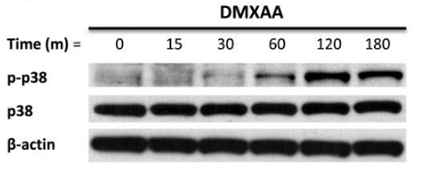
| Western blot | p-p38 / p38 p-MK2 / pERK / p-JNK |
|
21819972 |
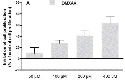
| Growth inhibition assay | Cell proliferation |
|
30138430 |
Informazioni sullo studio clinico
(dati da https://clinicaltrials.gov, aggiornato il 2024-05-22)
| Numero NCT | Reclutamento | Condizioni | Sponsor/Collaboratori | Data di inizio | Fasi |
|---|---|---|---|---|---|
| NCT00856336 | Completed | Refractory Tumors |
Antisoma Research |
May 2003 | Phase 1 |
| NCT00863733 | Completed | Solid Tumors |
Cancer Research UK|Cancer Society Auckland |
May 1996 | Phase 1 |
Supporto tecnico
Istruzioni per la manipolazione
Tel: +1-832-582-8158 Ext:3
Per qualsiasi altra domanda, si prega di lasciare un messaggio.

I prodotti sono solo per uso di ricerca. Non per uso umano. Non vendiamo a pazienti.
©Copyright 2013 Selleck Chemicals. Tutti i diritti riservati.