solo per uso di ricerca
Stattic Inibitore STAT3
N. Cat.S7024
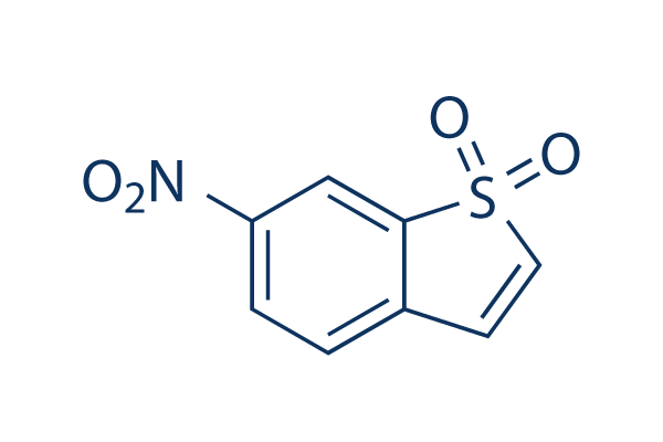
Struttura chimica
Peso molecolare: 211.19
Controllo Qualità
| Target correlati | EGFR JAK Pim |
|---|---|
| Altro STAT Inibitori | Napabucasin (BBI608) NSC 74859 (S3I-201) Cryptotanshinone (Tanshinone C) C188-9 (TTI-101) SH-4-54 BP-1-102 AS1517499 Nifuroxazide HO-3867 Homoharringtonine (HHT) |
Coltura cellulare, trattamento e concentrazione di lavoro
| Linee cellulari | Tipo di saggio | Concentrazione | Tempo di incubazione | Formulazione | Descrizione dellattività | PMID |
|---|---|---|---|---|---|---|
| H9c2 | Function Assay | 10 μM | 4 h | reverses the effects of IL-27 | 26339633 | |
| NPC | Function Assay | 0-7.5 µM | abolishes EMT-like molecular alterations, and cell migration and invasion induced by RKIP knockdown | 25915430 | ||
| HASMC | Function Assay | 1.25-5 μM | 20 min | DMSO | inhibits p-(Y)-STAT-1,3,5 signals | 25849622 |
| H9c2 | Function Assay | 2/10 μM | 2 h | DMSO | abrogates the cytoprotective effects of IL-27 against SH | 25820907 |
| A431 | Growth Inhibition Assay | 2 μM | 2 h | blocks EGF-reversed decreases in cell viability | 25720435 | |
| A431 | Growth Inhibition Assay | 2 μM | 2 h | increases in apoptosis induced by shikonin | 25720435 | |
| SiHa | Cell Viability Assay | 5-75 nM | 24 h | shows morphology of a typical apoptotic cell and dose-dependent loss of cell viability | 25539644 | |
| SiHa | Function Assay | 5-75 nM | 24 h | reduces the phosphorylation at the tyrosine residue 705 | 25539644 | |
| ECA109 | Growth Inhibition Assay | 0-20 μM | 24 h | IC50=5.50 μM | 25492480 | |
| TE13 | Growth Inhibition Assay | 0-20 μM | 24 h | IC50=6.15 μM | 25492480 | |
| KYSE150 | Growth Inhibition Assay | 0-20 μM | 24 h | IC50=12.64 μM | 25492480 | |
| ECA109 | Clonogenic Survival Assay | 0.5 μM | 24 h | suppresses the clonogenic formation | 25492480 | |
| TE13 | Clonogenic Survival Assay | 0.5 μM | 24 h | suppresses the clonogenic formation | 25492480 | |
| KYSE150 | Clonogenic Survival Assay | 0.5 μM | 24 h | suppresses the clonogenic formation | 25492480 | |
| ECA109 | Function Assay | 0.5 μM | 24 h | enhances IR-induced generation of DSBs | 25492480 | |
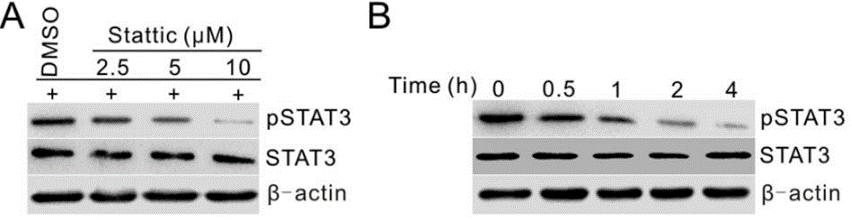
| PC3M-1E8 | Function Assay | 2.5/5/10 μM | 0-4 h | inhibits the STAT3 activation in a dose- and time-dependent manner | 25261365 | |
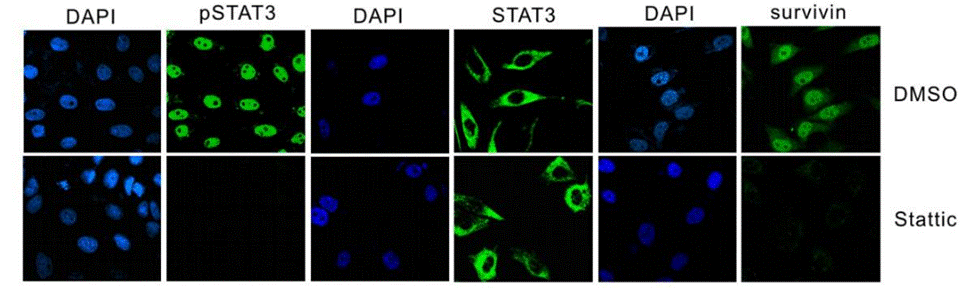
| PC3M-1E8 | Function Assay | 10 μM | 24 h | downregulates Bcl-xL, survivin and c-Myc | 25261365 | |
| PC3M-1E8 | Function Assay | 10 μM | 24 h | inhibits IL-6 induced STAT3 activation and the IL-6-induced STAT3 activation | 25261365 | |
| PC3M-1E8 | Clonogenic Survival Assay | 2.5/5/10 μM | inhibits the colony formation significantly | 25261365 | ||
| MDA-MB-231 | Function Assay | 20 μM | 2 h | exhibits Snail and E-cadherin expression | 25153349 | |
| H9c2 | Function Assay | 20 µM | 30 min | DMSO | abolishes propofol-induced AKT phosphorylation at both ser473 and thr308 | 25105067 |
| HaCaT | Growth Inhibition Assay | 10 µM | 20 min | DMSO | enhances sorafenib- and sunitinib-induced growth inhibition | 25013907 |
| Caki-1 | Growth Inhibition Assay | 10 µM | 20 min | DMSO | enhances sorafenib- and sunitinib-induced growth inhibition | 25013907 |
| HaCaT | Apoptosis Assay | 10 µM | 20 min | DMSO | increases proportions of apoptotic cells due to treatment with sorafenib or sunitinib | 25013907 |
| FHL-primed hNSCs | Cell Viability Assay | 0.02-5 μM | 72 h | leads to the loss of cell viability at high concentration | 24945434 | |
| ELL-primed hNSCs | Cell Viability Assay | 0.02-5 μM | 72 h | leads to the loss of cell viability at high concentration | 24945434 | |
| SS | Cell Viability Assay | 1-10 μM | 72 h | DMSO | causes a dose-dependent inhibition of the viability | 24756111 |
| SeAx | Cell Viability Assay | 1-10 μM | 72 h | DMSO | causes a dose-dependent inhibition of the viability | 24756111 |
| HuT-78 | Cell Viability Assay | 1-10 μM | 72 h | DMSO | causes a dose-dependent inhibition of the viability | 24756111 |
| CD4+ | Apoptosis Assay | 10 μm | 24 h | DMSO | induces apoptosis strongly | 24756111 |
| MCF-7 | Growth Inhibition Assay | 0.469-3.75 μM | 5 d | reduces cell number significantly | 24728078 | |
| MCF-7/LCC1 | Growth Inhibition Assay | 0.469-3.75 μM | 5 d | reduces cell number significantly | 24728078 | |
| MCF-7/LCC9 | Growth Inhibition Assay | 0.469-3.75 μM | 5 d | reduces cell number significantly | 24728078 | |
| HaCaT | Growth Inhibition Assay | 10 µM | 20 min | DMSO | enhances everolimus-induced cell growth inhibition | 24423131 |
| HaCaT | Apoptosis Assay | 10 µM | 20 min | DMSO | enhances the apoptotic effects of everolimus | 24423131 |
| MDA-MB-231 | Function Assay | 10 µM | 24 h | DMSO | reduces P-STAT3 expression | 24376586 |
| SUM-159 | Function Assay | 10 µM | 24 h | DMSO | reduces P-STAT3 expression | 24376586 |
| SK-BR-3 | Function Assay | 10 µM | 24 h | DMSO | reduces P-STAT3 expression | 24376586 |
| MCF7-HER2 | Growth Inhibition Assay | 0-10 μM | 48 h | DMSO | induces cell death dose dependently | 24297508 |
| MCF7-HER2 | Function Assay | 5 μM | 24 h | DMSO | diminishes Sox-2, Oct-4, and slug expression | 24297508 |
| MCF7-HER2 | Function Assay | 5 μM | 24 h | DMSO | decreases the expression levels of EMT markers, vimentin and slug | 24297508 |
| MCF7-HER2 | Growth Inhibition Assay | 5 μM | 24 h | DMSO | enhances cell growth inhibition combined with Herceptin | 24297508 |
| HMECs | Function Assay | 10 μM | 2 h | inhibits IFNα mediated phosphorylation of STAT1, STAT2 and STAT3 | 24211327 | |
| HTR8/SVneo | Function Assay | 1 μM | 1 h | suppressed OSM-induced STAT3 phosphorylation | 24060241 | |
| HTR8/SVneo | Function Assay | 0.5/1 μM | 48 h | restores the expression of E-cadherin suppressed by OSM | 24060241 | |
| HTR8/SVneo | Function Assay | 1 μM | 48 h | significantly increases migration by OSM | 24060241 | |
| C13* | Apoptosis Assay | 0-10 μM | 24/48 h | induces apoptosis in a dose and time dependent manner | 23962558 | |
| OV2008 | Apoptosis Assay | 0-10 μM | 24/48 h | induces apoptosis in a dose and time dependent manner | 23962558 | |
| C13* | Apoptosis Assay | 24/48 h | enhances cisplatin-induced apoptosis | 23962558 | ||
| OV2008 | Apoptosis Assay | 24/48 h | enhances cisplatin-induced apoptosis | 23962558 | ||
| W480 | Function Assay | 2.5/10 μM | 30 min | DMSO | sensitizes cells to chemoradiotherapy in a dose-dependent manner | 23934972 |
| SW837 | Function Assay | 2.5/10 μM | 30 min | DMSO | sensitizes cells to chemoradiotherapy in a dose-dependent manner | 23934972 |
| T24 | Function Assay | 2/10/20 μM | 24 h | causes dose-dependent inhibition of the CXCL12-induced increase of invading cells | 23526079 | |
| CNE1 | Function Assay | 20 µM | 48 h | blocks the IL-6 increased phosphorylation of Stat3 | 23382914 | |
| CNE2 | Function Assay | 20 µM | 48 h | blocks the IL-6 increased phosphorylation of Stat3 | 23382914 | |
| HONE1 | Function Assay | 20 µM | 48 h | blocks the IL-6 increased phosphorylation of Stat3 | 23382914 | |
| CNE1 | Growth Inhibition Assay | 4 μM | significantly reduces cell viability | 23382914 | ||
| CNE1 | Function Assay | 0-20 μM | 0-4 h | inhibits Stat3 activation in a dose- and time-dependent manner | 23382914 | |
| CNE2 | Function Assay | 0-20 μM | 0-4 h | inhibits Stat3 activation in a dose- and time-dependent manner | 23382914 | |
| HONE1 | Function Assay | 0-20 μM | 0-4 h | inhibits Stat3 activation in a dose- and time-dependent manner | 23382914 | |
| CNE1 | Cell Viability Assay | 0.5-64 μM | 48 h | suppresses cell viability in a dose- and time-dependent manner | 23382914 | |
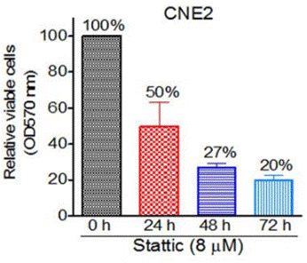
| CNE2 | Cell Viability Assay | 0.5-64 μM | 48 h | suppresses cell viability in a dose- and time-dependent manner | 23382914 | |
| HONE1 | Cell Viability Assay | 0.5-64 μM | 48 h | suppresses cell viability in a dose- and time-dependent manner | 23382914 | |
| C666-1 | Cell Viability Assay | 0.5-64 μM | 48 h | suppresses cell viability in a dose- and time-dependent manner | 23382914 | |
| CNE1 | Apoptosis Assay | 10 µM | 48 h | induces apoptosis | 23382914 | |
| CNE2 | Apoptosis Assay | 10 µM | 48 h | induces apoptosis | 23382914 | |
| HONE1 | Apoptosis Assay | 10 µM | 48 h | induces apoptosis | 23382914 | |
| CNE2 | Cell Viability Assay | 1/2 μM | 48 h | sensitize cells to radiotherapy | 23382914 | |
| HONE1 | Cell Viability Assay | 1/2 μM | 48 h | sensitize cells to radiotherapy | 23382914 | |
| C666-1 | Cell Viability Assay | 1/2 μM | 48 h | sensitize cells to radiotherapy | 23382914 | |
| HEC-1A | Function Assay | 1 μM | 24 h | DMSO | blocks the MUC20-enhanced invasion triggered by 10% FBS | 23262208 |
| RL95-2 | Function Assay | 1 μM | 24 h | DMSO | blocks the MUC20-enhanced invasion triggered by 10% FBS | 23262208 |
| HEC-1A | Function Assay | 1 μM | 24 h | DMSO | blocks the MUC20-enhanced invasion triggered by EGF | 23262208 |
| RL95-2 | Function Assay | 1 μM | 24 h | DMSO | blocks the MUC20-enhanced invasion triggered by EGF | 23262208 |
| CT26 | Function Assay | 20 mM | 1 h | suppresses HGF-induced VEGF expression | 23233163 | |
| UM-SCC-17B | Growth Inhibition Assay | IC50=2.562 ± 0.409 μM, GI50=1.279 ± 0.194 μM | 22770899 | |||
| OSC-19 | Growth Inhibition Assay | IC50=3.481 ± 0.953 μM, GI50=1.366 ± 0.770 μM | 22770899 | |||
| Cal33 | Growth Inhibition Assay | IC50=2.282 ± 0.423 μM, GI50=1.349 ± 0.363 μM | 22770899 | |||
| UM-SCC-22B | Growth Inhibition Assay | IC50=2.648 ± 0.542 μM, GI50=1.320 ± 0.204 μM | 22770899 | |||
| UM-SCC-17B | Function Assay | 0-30 μM | 0-24 h | inhibits STAT3 activation dose and time dependently | 22770899 | |
| OSC-19 | Function Assay | 0-30 μM | 0-24 h | inhibits STAT3 activation dose and time dependently | 22770899 | |
| Cal33 | Function Assay | 0-30 μM | 0-24 h | inhibits STAT3 activation dose and time dependently | 22770899 | |
| UM-SCC-22B | Function Assay | 0-30 μM | 0-24 h | inhibits STAT3 activation dose and time dependently | 22770899 | |
| U-87MG | Cell Viability Assay | 0-10 μM | 72 h | DMSO | inhibits cell viability dose dependently | 25436682 |
| U-373MG | Cell Viability Assay | 0-10 μM | 72 h | DMSO | inhibits cell viability dose dependently | 25436682 |
| SH-SY5Y | Cell Viability Assay | 0-10 μM | 72 h | DMSO | inhibits cell viability dose dependently | 25436682 |
| Tu-9648 | Cell Viability Assay | 0-10 μM | 72 h | DMSO | inhibits cell viability dose dependently | 25436682 |
| Neuro-2a | Cell Viability Assay | 0-10 μM | 72 h | DMSO | inhibits cell viability dose dependently | 25436682 |
| PCNs | Cell Viability Assay | 0-10 μM | 72 h | DMSO | inhibits cell viability dose dependently | 25436682 |
| PGCs | Cell Viability Assay | 0-10 μM | 72 h | DMSO | inhibits cell viability dose dependently | 25436682 |
| RAW264.7 | Function Assay | 10 μM | 12 h | abrogates the mRNA expressions of JAK2, STAT1, STAT2, and STAT3 induced by DON and T-2 toxin | 22454431 | |
| RAW264.7 | Apoptosis Assay | 5/10 μM | 45 min | enhances toxins induced apoptosis and MMP loss | 22454431 | |
| SW480 | Cell Viability Assay | 5/10/20 μM | 72 h | inhibits cell viability of the ALDH+/CD133+ cells | 21900397 | |
| HCT116 | Cell Viability Assay | 5/10/20 μM | 72 h | inhibits cell viability of the ALDH+/CD133+ cells | 21900397 | |
| DLD-1 | Cell Viability Assay | 5/10/20 μM | 72 h | inhibits cell viability of the ALDH+/CD133+ cells | 21900397 | |
| SNU387 | Cell Viability Assay | 20 μM | 24 h | reduces cell viability | 21311975 | |
| SNU398 | Cell Viability Assay | 20 μM | 24 h | reduces cell viability | 21311975 | |
| HepG2 | Cell Viability Assay | 20 μM | 24 h | reduces cell viability | 21311975 | |
| Huh-7 | Cell Viability Assay | 20 μM | 24 h | reduces cell viability | 21311975 | |
| VSMC | Growth Inhibition Assay | 3/5/10 μM | 30 min | DMSO | prevents PDGF- and thrombin-mediated VSMC proliferation in a dose-dependent manner | 20847306 |
| MDA-MB-231 | Apoptosis Assay | 10 μM | 24 h | DMSO | induces apoptosis | 17114005 |
| MDA-MB-435S | Apoptosis Assay | 10 μM | 24 h | DMSO | induces apoptosis | 17114005 |
| AsPC1 | Antiproliferative assay | 72 hrs | Antiproliferative activity against human AsPC1 cells assessed as inhibition of cell proliferation after 72 hrs by MTS assay, IC50 = 1.32 μM. | 24904966 | ||
| MDA-MB-231 | Antiproliferative assay | 72 hrs | Antiproliferative activity against ER-negative and triple-negative human MDA-MB-231 cells assessed as inhibition of cell proliferation after 72 hrs by MTS assay, IC50 = 2.89 μM. | 24904966 | ||
| MCF7 | Antiproliferative assay | 72 hrs | Antiproliferative activity against ER-positive human MCF7 cells assessed as inhibition of cell proliferation after 72 hrs by MTS assay, IC50 = 3.6 μM. | 24904966 | ||
| PANC1 | Antiproliferative assay | 72 hrs | Antiproliferative activity against human PANC1 cells assessed as inhibition of cell proliferation after 72 hrs by MTS assay, IC50 = 3.77 μM. | 24904966 | ||
| MDA-MB-231 | Cytotoxicity assay | 48 hrs | Cytotoxicity against human MDA-MB-231 cells assessed as growth inhibition after 48 hrs by MTT assay, IC50 = 1.56 μM. | 26396689 | ||
| MDA-MB-435S | Cytotoxicity assay | 48 hrs | Cytotoxicity against human MDA-MB-435S cells assessed as growth inhibition after 48 hrs by MTT assay, IC50 = 1.87 μM. | 26396689 | ||
| MCF7 | Cytotoxicity assay | 48 hrs | Cytotoxicity against human MCF7 cells assessed as growth inhibition after 48 hrs by MTT assay, IC50 = 2.16 μM. | 26396689 | ||
| A549 | Cytotoxicity assay | 48 hrs | Cytotoxicity against human A549 cells assessed as growth inhibition after 48 hrs by MTT assay, IC50 = 2.5 μM. | 26396689 | ||
| DU145 | Cytotoxicity assay | 48 hrs | Cytotoxicity against human DU145 cells assessed as growth inhibition after 48 hrs by MTT assay, IC50 = 2.5 μM. | 26396689 | ||
| PANC1 | Cytotoxicity assay | 48 hrs | Cytotoxicity against human PANC1 cells assessed as growth inhibition after 48 hrs by MTT assay, IC50 = 2.9 μM. | 26396689 | ||
| HCT116 | Antiproliferative assay | 72 hrs | Antiproliferative activity against human HCT116 cells after 72 hrs by MTT assay, IC50 = 1.08 μM. | 27718470 | ||
| MDA-MB-231 | Antiproliferative assay | 72 hrs | Antiproliferative activity against human MDA-MB-231 cells after 72 hrs by MTT assay, IC50 = 1.68 μM. | 27718470 | ||
| MCF7 | Antiproliferative assay | 72 hrs | Antiproliferative activity against human MCF7 cells after 72 hrs by MTT assay, IC50 = 2.36 μM. | 27718470 | ||
| A549 | Antiproliferative assay | 72 hrs | Antiproliferative activity against human A549 cells after 72 hrs by MTT assay, IC50 = 4.4 μM. | 27718470 | ||
| AD293 | Function assay | 6 hrs | Inhibition of IFNgamma-stimulated GFP/FLAG-tagged STAT3 dimerization in human AD293 cells incubated for 6 hrs by Western blot analysis, IC50 = 5.1 μM. | 30228000 | ||
| MDA-MB-231 | Function assay | 1 to 10 uM | 12 hrs | Inhibition of STAT3 phosphorylation at Tyr705 in human MDA-MB-231 cells at 1 to 10 uM after 12 hrs by western blot analysis | 24904966 | |
| MDA-MB-231 | Anticancer assay | 1 to 10 uM | 48 hrs | Anticancer activity against human MDA-MB-231 cells assessed as cell growth inhibition, apoptosis and cellular morphological changes at 1 to 10 uM after 48 hrs by light microscopy | 24904966 | |
| MDA-MB-231 | Function assay | 1 to 10 uM | 12 hrs | Decrease in STAT3 protein expression in human MDA-MB-231 cells at 1 to 10 uM after 12 hrs by western blot analysis | 24904966 | |
| MCF7 | Function assay | 12 hrs | Inhibition of STAT3 phosphorylation at Y705 in human MCF7 cells after 12 hrs by Western blot analysis | 26396689 | ||
| MDA-MB-435S | Function assay | 12 hrs | Inhibition of STAT3 phosphorylation at Y705 in human MDA-MB-435S cells after 12 hrs by Western blot analysis | 26396689 | ||
| MDA-MB-231 | Function assay | 12 hrs | Inhibition of STAT3 phosphorylation at Y705 in human MDA-MB-231 cells after 12 hrs by Western blot analysis | 26396689 | ||
| Clicca per visualizzare più dati sperimentali sulle linee cellulari | ||||||
Informazioni chimiche, conservazione e stabilità
| Peso molecolare | 211.19 | Formula | C8H5NO4S |
Conservazione (Dalla data di ricezione) | |
|---|---|---|---|---|---|
| N. CAS | 19983-44-9 | Scarica SDF | Conservazione delle soluzioni stock |
|
|
| Sinonimi | N/A | Smiles | C1=CC(=CC2=C1C=CS2(=O)=O)[N+](=O)[O-] | ||
Solubilità
|
In vitro |
DMSO
: 42 mg/mL
(198.87 mM)
Water : Insoluble Ethanol : Insoluble |
Calcolatore di Molarità
|
In vivo |
|||||
Calcolatore di formulazione in vivo (Soluzione chiara)
Passo 1: Inserire le informazioni di seguito (Consigliato: Un animale aggiuntivo per tenere conto della perdita durante lesperimento)
Passo 2: Inserire la formulazione in vivo (Questo è solo il calcolatore, non la formulazione. Contattateci prima se non cè una formulazione in vivo nella sezione Solubilità.)
Risultati del calcolo:
Concentrazione di lavoro: mg/ml;
Metodo per preparare il liquido master di DMSO: mg farmaco predissolto in μL DMSO ( Concentrazione del liquido master mg/mL, Vi preghiamo di contattarci prima se la concentrazione supera la solubilità del DMSO del lotto del farmaco. )
Metodo per preparare la formulazione in vivo: Prendere μL DMSO liquido master, quindi aggiungereμL PEG300, mescolare e chiarire, quindi aggiungereμL Tween 80, mescolare e chiarire, quindi aggiungere μL ddH2O, mescolare e chiarire.
Metodo per preparare la formulazione in vivo: Prendere μL DMSO liquido master, quindi aggiungere μL Olio di mais, mescolare e chiarire.
Nota: 1. Si prega di assicurarsi che il liquido sia limpido prima di aggiungere il solvente successivo.
2. Assicurarsi di aggiungere il/i solvente/i in ordine. È necessario assicurarsi che la soluzione ottenuta, nellaggiunta precedente, sia una soluzione limpida prima di procedere allaggiunta del solvente successivo. Metodi fisici come il vortex, gli ultrasuoni o il bagno dacqua calda possono essere utilizzati per facilitare la dissoluzione.
Meccanismo dazione
| Caratteristiche |
Stattic is the first non-peptide small molecule with inhibitory activity against STAT3 SH2 domain regardless of the STAT3 phosphorylation state in vitro.
|
|---|---|
| Targets/IC50/Ki |
STAT3
(Cell-free assay) 5.1 μM
|
| In vitro |
Stattic inibisce il legame di un peptide contenente fosfotirosina derivato dal recettore gp130 al dominio SH2 di STAT3 in modo fortemente dipendente dalla temperatura. Questo composto ha solo un effetto molto debole sul legame di un peptide tirosina-fosforilato al dominio SH2 della tirosina chinasi Lck. E non inibisce la dimerizzazione di altri due fattori di trascrizione dimerici (c-Myc/Max e Jun/Jun). Inibisce anche i fosfopeptidi marcati con fluoresceina ai domini SH2 di STAT1 e STAT5b. Questa sostanza chimica inibisce selettivamente il legame del DNA degli omodimeri STAT3 a una concentrazione di 10 μM. È stato dimostrato che inibisce la fosforilazione cellulare di STAT3 a Tyr705 con scarso effetto sulla fosforilazione di STAT1 a Tyr701 (nelle cellule HepG2) o sulla fosforilazione di JAK1, JAK2 e c-Src (nelle cellule MDA-MB-231 e MDA-MB-235S). Questo composto aumenta il tasso apoptotico delle linee cellulari di cancro al seno dipendenti da STAT3. |
| Saggio chinasico |
Screening ad alto rendimento e saggi di polarizzazione della fluorescenza
|
|
Lo screening viene eseguito a circa 30 °C. La specificità dei risultati dello screening viene convalidata in saggi analoghi per il legame dei composti di prova ai domini SH2 di STAT1, STAT5 e Lck. La concentrazione finale dei componenti del tampone utilizzati per tutti i saggi FP è 10 mM HEPES (pH 7,5), 1 mM EDTA, 0,1% Nonidet P-40, 50 mM NaCl e 10% DMSO. L'assenza di ditiotreitolo è essenziale per l'attività inibitoria. Le sequenze dei peptidi sono: STAT3, 5-carbossifluoresceina-GY(PO3H2)LPQTV-NH2; STAT1, 5-carbossifluoresceina-GY(PO3H2)DKPHVL; STAT5, 5-carbossifluoresceina-GY(PO3H2)LVLDKW; e Lck, 5-carbossifluoresceina-GY(PO3H2)EEIP. Per l'analisi di specificità a 30 °C, le proteine vengono utilizzate a 150 nM (STAT1, STAT3 e STAT5). Per l'analisi di specificità a 37 °C, le proteine vengono utilizzate a 370 nM (STAT3) o 100 nM (Lck). Le proteine vengono incubate con i composti di prova in provette Eppendorf alle temperature indicate per 60 minuti prima dell'aggiunta dei rispettivi peptidi marcati con 5-carbossifluoresceina (concentrazione finale: 10 nM). Prima della misurazione a temperatura ambiente, le miscele vengono lasciate equilibrare per almeno 30 minuti. I composti di prova vengono utilizzati alle concentrazioni indicate diluite da una soluzione madre 20× in DMSO. Le curve di legame e le curve di inibizione vengono adattate con SigmaPlot. Tutte le curve di competizione vengono ripetute tre volte in esperimenti indipendenti.
|
Riferimenti |
|
Applicazioni
| Metodi | Biomarcatori | Immagini | PMID |
|---|---|---|---|
| Western blot | p-STAT3 / STAT3 Survivin / c-Myc / Bcl-xl PARP / C-PARP / Caspase-3 / C-Caspse-3 |
|
25261365 |
| Immunofluorescence | p-STAT3 / STAT3 / Survivin |
|
25261365 |
| Growth inhibition assay | Cell viability |
|
23382914 |
| ELISA | BDNF |
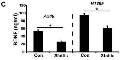
|
27456333 |
Supporto tecnico
Istruzioni per la manipolazione
Tel: +1-832-582-8158 Ext:3
Per qualsiasi altra domanda, si prega di lasciare un messaggio.
I prodotti sono solo per uso di ricerca. Non per uso umano. Non vendiamo a pazienti.
©Copyright 2013 Selleck Chemicals. Tutti i diritti riservati.