solo per uso di ricerca
NSC 74859 (S3I-201) Inibitore di STAT3
N. Cat.S1155
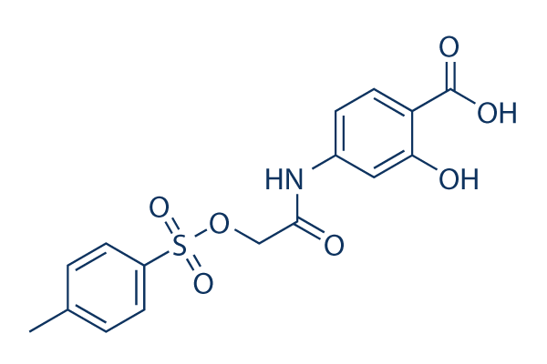
Struttura chimica
Peso molecolare: 365.36
Controllo Qualità
| Target correlati | EGFR JAK Pim |
|---|---|
| Altro STAT Inibitori | Napabucasin (BBI608) Stattic Cryptotanshinone (Tanshinone C) C188-9 (TTI-101) SH-4-54 BP-1-102 AS1517499 Nifuroxazide HO-3867 Homoharringtonine (HHT) |
Coltura cellulare, trattamento e concentrazione di lavoro
| Linee cellulari | Tipo di saggio | Concentrazione | Tempo di incubazione | Formulazione | Descrizione dellattività | PMID |
|---|---|---|---|---|---|---|
| U87 | Growth Inhibition Assay | 72 h | IC50=55.1 μM | 20072652 | ||
| U373 | Growth Inhibition Assay | 72 h | IC50=52.5 μM | 20072652 | ||
| HPAC | Growth Inhibition Assay | 72 h | IC50>100 μM | 20072652 | ||
| PANC-1 | Growth Inhibition Assay | 72 h | IC50>100 μM | 20072652 | ||
| SK-BR-3 | Growth Inhibition Assay | 72 h | IC50>100 μM | 20072652 | ||
| U-373 MG | Cytotoxicity Assay | 3/10 μM | 24 h | reduces FN-γ-induced cell neurotoxicity | 20888416 | |
| MDA-MB-231 | Growth Inhibition Assay | 72 h | IC50>100 μM | 20072652 | ||
| HUVEC | Function Assay | 0.5-20 μM | 24 h | DMSO | suppresses the hypoxia-induced accumulation of HIF-1α | 21523559 |
| Huh7 | Growth Inhibition Assay | 100 nM | 48 h | DMSO | inhibits the IL-6 stimulation promoted cell proliferation | 23364389 |
| PLC/PRF/5 | Growth Inhibition Assay | 100 nM | 48 h | DMSO | inhibits the IL-6 stimulation promoted cell proliferation | 23364389 |
| H460 | Function Assay | 50/100 μM | 48 h | inhibits the Stat3C increased miR-92a expression | 23820254 | |
| H1299 | Function Assay | 50/100 μM | 48 h | suppresses miR-92a expression dose-dependently | 23820254 | |
| T-cell | Growth Inhibition Assay | IC50=50 μM | 24068731 | |||
| U373 | Growth Inhibition Assay | 125 μM | 24 h | DMSO | disrupts STAT3 signaling and proliferation | 24070820 |
| HUT-102 | Apoptosis Assay | 75-300 μM | 24/48 h | suppresses cell proliferation in a dose-dependent manner and induces cell apoptosis | 24090995 | |
| MT-2 | Apoptosis Assay | 75-300 μM | 24/48 h | suppresses cell proliferation in a dose-dependent manner and induces cell apoptosis | 24090995 | |
| H460 | Apoptosis Assay | 100 nM | 24 h | enhances cell death co-treated with LY294002 | 24472538 | |
| A459 | Apoptosis Assay | 100 nM | 24 h | induces cell apoptosis co-treated with BEZ235 | 24472538 | |
| H460 | Apoptosis Assay | 100 nM | 24 h | induces cell apoptosis co-treated with BEZ235 | 24472538 | |
| GC | Growth Inhibition Assay | 50-125 μM | 72 h | attenuates the cell growth in a dose-dependent manner | 25774503 | |
| GH3 | Growth Inhibition Assay | 50-125 μM | 72 h | attenuates the cell growth in a dose-dependent manner | 25774503 | |
| BT474R | Function Assay | 50 μM | 10-60 d | inhibits STAT3 activity | 25327561 | |
| NCI-N87R | Function Assay | 50 μM | 10-60 d | inhibits STAT3 activity | 25327561 | |
| MDA-MB-468 | Function assay | 100 uM | 24 hrs | Inhibition of Stat3 activation in human MDA-MB-468 cells at 100 uM after 24 hrs | 17463090 | |
| MDA-MB-435 | Function assay | 100 uM | 24 hrs | Inhibition of Stat3 activation in human MDA-MB-435 cells at 100 uM after 24 hrs | 17463090 | |
| MDA-MB-231 | Function assay | 100 uM | 24 hrs | Inhibition of Stat3 activation in human MDA-MB-231 cells at 100 uM after 24 hrs | 17463090 | |
| NIH3T3 | Function assay | 100 uM | 24 hrs | Reduction of pTyr-705 Stat3 level in v-Src expressing mouse NIH3T3 cells at 100 uM after 24 hrs | 17463090 | |
| NIH3T3 | Growth inhibition assay | 100 uM | 4 days | Growth inhibition of mouse NIH3T3 cells expressing v-Src at 100 uM after 4 days by trypan blue exclusion assay | 17463090 | |
| MDA-MB-435 | Growth inhibition assay | 100 uM | 4 days | Growth inhibition of human MDA-MB-435 cells expressing v-Src at 100 uM after 4 days by trypan blue exclusion assay | 17463090 | |
| MDA-MB-231 | Growth inhibition assay | 100 uM | 4 days | Growth inhibition of human MDA-MB-231 cells expressing v-Src at 100 uM after 4 days by trypan blue exclusion assay | 17463090 | |
| MDA-MB-468 | Growth inhibition assay | 100 uM | 4 days | Growth inhibition of human MDA-MB-468 cells expressing v-Src at 100 uM after 4 days by trypan blue exclusion assay | 17463090 | |
| NIH3T3 | Growth inhibition assay | 100 uM | Growth inhibition of mouse NIH3T3 cells expressing v-Ras at 100 uM for every 3 days by soft-agar colony-formation assay | 17463090 | ||
| MDA-MB-435 | Apoptosis assay | 30 to 100 uM | 48 hrs | Induction of apoptosis in human MDA-MB-435 cells expressing active Stat3 at 30 to 100 uM after 48 hrs | 17463090 | |
| MDA-MB-231 | Apoptosis assay | 100 uM | 24 hrs | Reduction of apoptosis in Stat3 transfected human MDA-MB-231 cells at 100 uM after 24 hrs | 17463090 | |
| MDA-MB-231 | Function assay | 100 uM | 48 hrs | Reduction of cyclin D1 gene expression in human MDA-MB-231 cells at 100 uM after 48 hrs | 17463090 | |
| MDA-MB-231 | Apoptosis assay | 100 uM | 24 hrs | Induction of apoptosis in Stat3 SH2 domain transfected human MDA-MB-231 cells at 100 uM after 24 hrs | 17463090 | |
| MDA-MB-231 | Apoptosis assay | 100 uM | 24 hrs | Induction of apoptosis in Stat3C transfected human MDA-MB-231 cells at 100 uM after 24 hrs | 17463090 | |
| NIH3T3 | Function assay | 100 uM | 48 hrs | Reduction of cyclin D1 gene expression in v-Src transfected mouse NIH3T3 cells at 100 uM after 48 hrs | 17463090 | |
| NIH3T3 | Function assay | 100 uM | 48 hrs | Reduction of Bcl-xL gene expression in v-Src transfected mouse NIH3T3 cells at 100 uM after 48 hrs | 17463090 | |
| NIH3T3 | Function assay | 100 uM | 48 hrs | Reduction of survivin gene expression in v-Src transfected mouse NIH3T3 cells at 100 uM after 48 hrs | 17463090 | |
| MDA-MB-231 | Function assay | 100 uM | 48 hrs | Reduction of Bcl-xL gene expression in human MDA-MB-231 cells at 100 uM after 48 hrs | 17463090 | |
| MDA-MB-231 | Function assay | 100 uM | 48 hrs | Reduction of survivin gene expression in human MDA-MB-231 cells at 100 uM after 48 hrs | 17463090 | |
| MDA-MB-231 | Antitumor assay | 5 mg/kg | 2 weeks | Antitumor activity against human MDA-MB-231 cells expressing active Stat3 xenografted in mouse at 5 mg/kg, iv for every 3 days for 2 weeks | 17463090 | |
| NIH3T3 | Growth inhibition assay | 100 uM | Growth inhibition of mouse NIH3T3 cells expressing v-Src at 100 uM for every 3 days by soft-agar colony-formation assay | 17463090 | ||
| A673 | qHTS assay | qHTS of pediatric cancer cell lines to identify multiple opportunities for drug repurposing: Primary screen for A673 cells | 29435139 | |||
| SK-N-MC | qHTS assay | qHTS of pediatric cancer cell lines to identify multiple opportunities for drug repurposing: Primary screen for SK-N-MC cells | 29435139 | |||
| NB1643 | qHTS assay | qHTS of pediatric cancer cell lines to identify multiple opportunities for drug repurposing: Primary screen for NB1643 cells | 29435139 | |||
| LAN-5 | qHTS assay | qHTS of pediatric cancer cell lines to identify multiple opportunities for drug repurposing: Primary screen for LAN-5 cells | 29435139 | |||
| Clicca per visualizzare più dati sperimentali sulle linee cellulari | ||||||
Informazioni chimiche, conservazione e stabilità
| Peso molecolare | 365.36 | Formula | C16H15NO7S |
Conservazione (Dalla data di ricezione) | |
|---|---|---|---|---|---|
| N. CAS | 501919-59-1 | Scarica SDF | Conservazione delle soluzioni stock |
|
|
Solubilità
|
In vitro |
DMSO
: 73 mg/mL
(199.8 mM)
Water : Insoluble Ethanol : Insoluble |
Calcolatore di Molarità
|
In vivo |
|||||
Calcolatore di formulazione in vivo (Soluzione chiara)
Passo 1: Inserire le informazioni di seguito (Consigliato: Un animale aggiuntivo per tenere conto della perdita durante lesperimento)
Passo 2: Inserire la formulazione in vivo (Questo è solo il calcolatore, non la formulazione. Contattateci prima se non cè una formulazione in vivo nella sezione Solubilità.)
Risultati del calcolo:
Concentrazione di lavoro: mg/ml;
Metodo per preparare il liquido master di DMSO: mg farmaco predissolto in μL DMSO ( Concentrazione del liquido master mg/mL, Vi preghiamo di contattarci prima se la concentrazione supera la solubilità del DMSO del lotto del farmaco. )
Metodo per preparare la formulazione in vivo: Prendere μL DMSO liquido master, quindi aggiungereμL PEG300, mescolare e chiarire, quindi aggiungereμL Tween 80, mescolare e chiarire, quindi aggiungere μL ddH2O, mescolare e chiarire.
Metodo per preparare la formulazione in vivo: Prendere μL DMSO liquido master, quindi aggiungere μL Olio di mais, mescolare e chiarire.
Nota: 1. Si prega di assicurarsi che il liquido sia limpido prima di aggiungere il solvente successivo.
2. Assicurarsi di aggiungere il/i solvente/i in ordine. È necessario assicurarsi che la soluzione ottenuta, nellaggiunta precedente, sia una soluzione limpida prima di procedere allaggiunta del solvente successivo. Metodi fisici come il vortex, gli ultrasuoni o il bagno dacqua calda possono essere utilizzati per facilitare la dissoluzione.
Meccanismo dazione
| Caratteristiche |
A chemical probe inhibitor of Stat3 activity.
|
|---|---|
| Targets/IC50/Ki |
STAT3
(Cell-free assay) 86 μM
|
| In vitro |
NSC 74859 (S3I-201) inibisce la crescita e induce l'apoptosi preferenzialmente nelle cellule tumorali che contengono Stat3 persistentemente attivata, inibendo la formazione del complesso Stat3·Stat3 e le attività di legame al DNA e trascrizionali di Stat3. Inoltre, inibisce anche l'espressione dei geni regolati da Stat3 che codificano per ciclina D1, Bcl-xL e survivina. Questo composto inibisce le linee cellulari di carcinoma mammario MDA-MB-435, MDA-MB-453 e MDA-MB-231 con IC50 di 100 μM. Inoltre, le cellule con segnalazione TGF-β compromessa sono quattro volte più sensibili a S3I-201. Uno studio recente dimostra che potenzia l'effetto antiproliferativo nelle cellule HepG2 e Huh-7 tramite la via di segnalazione STAT3. |
| Saggio chinasico |
Saggio di legame al DNA di Stat3 in vitro e analisi EMSA
|
|
In breve, 100 μL di biotinil-e-Ac-EPQpYEEIEL-OH (in 50 mM Tris/150 mM NaCl, pH 7.5) vengono aggiunti a ciascun pozzetto di piastre per microtitolazione a 96 pozzetti rivestite con streptavidina e incubati con agitazione a 4 °C durante la notte. Le piastre vengono quindi risciacquate con PBS/Tween 20 e quindi due volte con 200 μL di BSA-T-PBS (0.2% BSA/0.1% Tween 20/PBS). Quindi, 50 μL di proteina di fusione Lck-SH2-GST (6.4 ng/ml in BSA-T-PBS) vengono aggiunti a ciascun pozzetto della piastra a 96 pozzetti in presenza e assenza di 50 μL di NSC 74859 (S3I-201) (per concentrazioni finali di 30 e 100 mM), e la piastra viene agitata a temperatura ambiente per 4 ore. Dopo aver rimosso le soluzioni, ciascun pozzetto viene risciacquato quattro volte con BSA-T-PBS (200 μL), e 100 μL di anticorpo policlonale di coniglio anti-GST (100 ng/mL in BSA-T-PBS) vengono aggiunti a ciascun pozzetto e incubati a 4 °C durante la notte. Dopo il lavaggio con BSA-T-PBS, 100 μL di anticorpo di topo anti-coniglio coniugato con perossidasi di rafano BSA-T-PBS da 200 ng/mL vengono aggiunti a ciascun pozzetto e incubati per 45 minuti a temperatura ambiente. Dopo quattro passaggi di lavaggio con BSA-T-PBS e tre passaggi di lavaggio con PBS-T, 100 μL di substrato perossidasico vengono aggiunti a ciascun pozzetto e incubati per 5-15 minuti. La reazione perossidasica viene interrotta aggiungendo 100 μL di soluzione di acido solforico 1 M, e l'assorbanza viene letta a 450 nm con un lettore di piastre ELISA.
|
|
| In vivo |
NSC 74859 (S3I-201) (5 mg/kg, i.v. ogni 2 o ogni 3 giorni) mostra l'efficacia antitumorale in modelli murini con xenotrapianti di tumori mammari umani che ospitano Stat3 costitutivamente attiva. Questo trattamento con il composto riduce la replicazione del virus Varicella-zoster (VZV) sulla base del segnale di bioluminescenza e del numero di xenotrapianti cutanei positivi rispetto ai topi trattati con DMSO, inibendo la fosforilazione di STAT3. |
Riferimenti |
|
Applicazioni
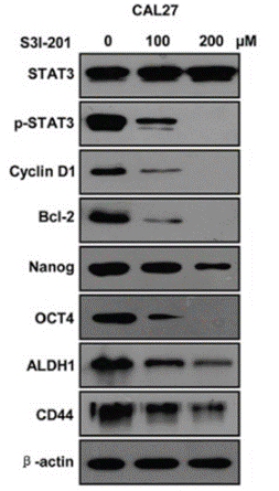
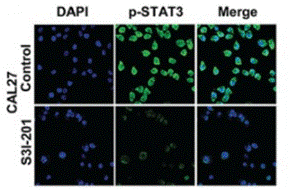
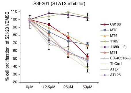
| Metodi | Biomarcatori | Immagini | PMID |
|---|---|---|---|
| Western blot | STAT3 / p-STAT3 / Cyclin D1 / Bcl-2 / Nanog / OCT4 / ALDH1 / CD44 PD-L1 |
|
26556875 |
| Immunofluorescence | p-STAT3 Oct4 / Twist |
|
26556875 |
| Growth inhibition assay | Cell viability |
|
26813676 |
Supporto tecnico
Istruzioni per la manipolazione
Tel: +1-832-582-8158 Ext:3
Per qualsiasi altra domanda, si prega di lasciare un messaggio.
I prodotti sono solo per uso di ricerca. Non per uso umano. Non vendiamo a pazienti.
©Copyright 2013 Selleck Chemicals. Tutti i diritti riservati.