solo per uso di ricerca
Monocyte + Macrophage Antibody [E17C4]
N. Cat.: F3741
-
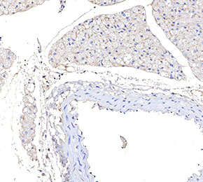 Immunohistochemical analysis of formalin fixed paraffin embedded mouse carotid artery tissue with F3741 at 1:100 dilution.
Elementi essenziali dellesperimento
This antibody requires an anti-rat secondary antibody.
Informazioni sullutilizzo
| Diluizione |
|---|
|
|
| Applicazione |
|---|
| IHC, FCM |
| Reattività |
|---|
| Mouse |
| Fonte |
|---|
| Rat Monoclonal Antibody |
| Tampone di conservazione |
|---|
| PBS, pH 7.2+50% Glycerol+0.05% BSA+0.01% NaN3 |
| Conservazione (dalla data di ricezione) |
|---|
| -20°C (avoid freeze-thaw cycles), 2 years |
Metodi sperimentali
| IHC |
|---|
Experimental Protocol:
Deparaffinization/Rehydration
1. Deparaffinize/hydrate sections:
2. Incubate sections in three washes of xylene for 5 min each.
3. Incubate sections in two washes of 100% ethanol for 10 min each.
4. Incubate sections in two washes of 95% ethanol for 10 min each.
5. Wash sections two times in dH2O for 5 min each.
6.Antigen retrieval: For Citrate: Heat slides in a microwave submersed in 1X citrate unmasking solution until boiling is initiated; continue with 10 min at a sub-boiling temperature (95°-98°C). Cool slides on bench top for 30 min.
Staining
1. Wash sections in dH2O three times for 5 min each.
2. Incubate sections in 3% hydrogen peroxide for 10 min.
3. Wash sections in dH2O two times for 5 min each.
4. Wash sections in wash buffer for 5 min.
5. Block each section with 100–400 µl of blocking solution for 1 hr at room temperature.
6. Remove blocking solution and add 100–400 µl primary antibody diluent in to each section. Incubate overnight at 4°C.
7. Remove antibody solution and wash sections with wash buffer three times for 5 min each.
8. Cover section with 1–3 drops HRPas needed. Incubate in a humidified chamber for 30 min at room temperature.
9. Wash sections three times with wash buffer for 5 min each.
10. Add DAB Chromogen Concentrate to DAB Diluent and mix well before use.
11. Apply 100–400 µl DAB to each section and monitor closely. 1–10 min generally provides an acceptable staining intensity.
12. Immerse slides in dH2O.
13. If desired, counterstain sections with hematoxylin.
14. Wash sections in dH2O two times for 5 min each.
15. Dehydrate sections: Incubate sections in 95% ethanol two times for 10 sec each; Repeat in 100% ethanol, incubating sections two times for 10 sec each; Repeat in xylene, incubating sections two times for 10 sec each.
16. Mount sections with coverslips and mounting medium.
|
Descrizione biologica
| Specificità |
|---|
Monocyte + Macrophage Antibody [E17C4] detects endogenous levels of total Monocyte + Macrophage antigen. This antibody recognises an intracellular antigen of mouse macrophages and monocytes. It reacts strongly with macrophages in lymphoid organs in all mouse strains. |
| Clone |
|---|
| E17C4 |
| Background |
|---|
| Monocytes and macrophages are essential components of the innate immune system, playing a central role in initiating and coordinating inflammatory responses. Beyond driving the production of inflammatory mediators and shaping both innate and adaptive immunity, they are equally critical in resolving inflammation and restoring tissue homeostasis. Dysregulation of their function is therefore a common feature in the pathogenesis of chronic infections, autoimmune conditions, and severe sterile inflammatory diseases. Monocytes, which represent 5–10% of circulating leukocytes, are bone marrow–derived mononuclear cells with a short life span of 1–3 days. Under steady-state conditions, they support homeostasis and retain the capacity to differentiate into tissue macrophages. During inflammation, monocytes are actively recruited to affected sites, where they differentiate into inflammatory macrophages or dendritic cells. Macrophages are distributed throughout all tissues of the body and display remarkable functional heterogeneity. They are indispensable for tissue development, immune surveillance, and the maintenance of local homeostasis. Many macrophage populations are established during embryogenesis, arising from yolk sac or fetal liver progenitors, and can persist independently of monocyte replenishment under normal conditions. By contrast, macrophages in tissues such as the skin, heart, and intestine are initially seeded by embryonic progenitors but are rapidly replaced after birth by monocytes derived from hematopoietic stem cells. Importantly, tissue-resident macrophages possess both proliferative capacity and self-renewal potential. The activation and polarization of both monocytes and macrophages are triggered by their recognition of pathogen-associated or damage-associated molecular patterns (PAMPs and DAMPs) via pattern recognition receptors (PRRs), enabling them to adapt their responses to a wide variety of physiological and pathological signals. |
| Riferimenti |
|---|
|
Supporto tecnico
Istruzioni per la manipolazione
Tel: +1-832-582-8158 Ext:3
Per qualsiasi altra domanda, si prega di lasciare un messaggio.
I prodotti sono solo per uso di ricerca. Non per uso umano. Non vendiamo a pazienti.
©Copyright 2013 Selleck Chemicals. Tutti i diritti riservati.