solo per uso di ricerca
Myosin Light Chain 2 Antibody [J14M12]
N. Cat.: F0485
-
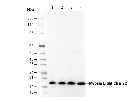 Lane 1: C2C12
Lane 2: A431
Lane 3: T24
Lane 4: 293T
Elementi essenziali dellesperimento
Recommended wet transfer conditions: 200 mA, 60 min,Recommended to use 0.22 μm PVDF membrane.
Informazioni sullutilizzo
| Diluizione |
|---|
|
| Applicazione |
|---|
| WB |
| Reattività |
|---|
| Human, Mouse, Rat |
| Fonte |
|---|
| Rabbit Monoclonal Antibody |
| Tampone di conservazione |
|---|
| PBS, pH 7.2+50% Glycerol+0.05% BSA+0.01% NaN₃ |
| Conservazione (dalla data di ricezione) |
|---|
| –20°C (avoid freeze-thaw cycles), 2 years |
| PM previsto |
|---|
| 18 kda |
| Controllo positivo | C2C12; A431; T24 |
|---|---|
| Controllo negativo |
Metodi sperimentali
| WB |
|---|
Experimental Protocol:
Sample preparation
1. Tissue: Lyse the tissue sample by adding an appropriate volume of ice-cold RIPA/Tris-Triton Lysis Buffer (containing Protease Inhibitor Cocktail),and homogenize the tissue at a low temperature or lyse it by sonication on ice, then incubate on ice for 30 minutes. 2. Adherent cell: Aspirate the culture medium and transfer the cells into an EP tube. Wash the cells with ice-cold PBS twice. Add an appropriate volume of RIPA/Tris-Triton Lysis Buffer (containing Protease Inhibitor Cocktail), sonicate to lyse the cells, and incubate on ice for 30 minutes. 3. Suspension cell: Transfer the culture medium to a pre-cooled centrifuge tube. Centrifuge and aspirate the supernatant. Wash the cells with ice-cold PBS twice.Add an appropriate volume of RIPA/Tris-Triton Lysis Buffer (containing Protease Inhibitor Cocktail), sonicate to lyse the cells, and incubate on ice for 30 minutes. 4. Place the lysate into a pre-cooled microcentrifuge tube. Centrifuge at 4°C for 15 min. Collect the supernatant;
5. Remove a small volume of lysate to determine the protein concentration;
6. Combine the lysate with protein loading buffer. Boil 20 µL sample under 95-100°C for 5 min. Centrifuge for 5 min after cool down on ice.
Electrophoretic separation
1. According to the concentration of extracted protein, load appropriate amount of protein sample and marker onto SDS-PAGE gels for electrophoresis. Recommended separating gel (lower gel) concentration: 10%. Reference Table for Selecting SDS-PAGE Separation Gel Concentrations 2. Power up 80V for 30 minutes. Then the power supply is adjusted (110 V~150 V), the Marker is observed, and the electrophoresis can be stopped when the indicator band of the predyed protein Marker where the protein is located is properly separated. (Note that the current should not be too large when electrophoresis, too large current (more than 150 mA) will cause the temperature to rise, affecting the result of running glue. If high currents cannot be avoided, an ice bath can be used to cool the bath.)
Transfer membrane
1. Take out the converter, soak the clip and consumables in the pre-cooled converter;
2. Activate PVDF membrane with methanol for 1 min and rinse with transfer buffer;
3. Install it in the order of "black edge of clip - sponge - filter paper - filter paper - glue -PVDF membrane - filter paper - filter paper - sponge - white edge of clip"; 4. The protein was electrotransferred to PVDF membrane. ( 0.22 µm PVDF membrane is recommended )) Reference Table for Selecting PVDF Membrane Pore Size Specifications Recommended conditions for wet transfer: 200 mA, 60 min. ( Note that the transfer conditions can be adjusted according to the protein size. For high-molecular-weight proteins, a higher current and longer transfer time are recommended. However, ensure that the transfer tank remains at a low temperature to prevent gel melting.)
Block
1. After electrotransfer, wash the film with TBST at room temperature for 5 minutes;
2. Incubate the film in the blocking solution for 1 hour at room temperature;
3. Wash the film with TBST for 3 times, 5 minutes each time.
Antibody incubation
1. Use 5% skim milk powder to prepare the primary antibody working liquid (recommended dilution ratio for primary antibody 1:1000), gently shake and incubate with the film at 4°C overnight; 2. Wash the film with TBST 3 times, 5 minutes each time;
3. Add the secondary antibody to the blocking solution and incubate with the film gently at room temperature for 1 hour;
4. After incubation, wash the film with TBST 3 times for 5 minutes each time.
Antibody staining
711. Add the prepared ECL luminescent substrate (or select other color developing substrate according to the second antibody) and mix evenly;
2. Incubate with the film for 1 minute, remove excess substrate (keep the film moist), wrap with plastic film, and expose in the imaging system. |
Descrizione biologica
| Specificità |
|---|
Myosin Light Chain 2 Antibody [J14M12] recognizes endogenous levels of total myosin light chain 2 protein. |
| Localizzazione subcellulare |
|---|
| Cytoplasm, Cytoskeleton |
| Uniprot ID |
|---|
| P24844 |
| Clone |
|---|
| J14M12 |
| Sinonimo |
|---|
| MYL9,Myosin Light Chain 2 |
| Background |
|---|
Myosin is made up of six polypeptide chains: two identical heavy chains and two pairs of light chains. Myosin light chain 2 (MLC2), also known as myosin regulatory light chain (MRLC), RLC, or LC20, has several isoforms based on its distribution. Myosin light chain-2 (MYL2, also called MLC-2) is a ∼19 kDa sarcomeric protein belonging to the EF-hand calcium-binding protein superfamily, existing in three major isoforms encoded by three distinct genes in mammalian striated muscle. These isoforms are: MLC-2f (fast twitch skeletal isoform), MLC-2v (cardiac ventricular and slow twitch skeletal isoform), and MLC-2a (cardiac atrial isoform), each with a unique developmental expression pattern in mammals. In smooth muscle, MLC2 is phosphorylated at Thr18 and Ser19 by myosin light chain kinase (MLCK) in a Ca2+/calmodulin-dependent manner, which is associated with myosin ATPase activity and smooth muscle contraction. |
| Riferimenti |
|---|
|
Supporto tecnico
Istruzioni per la manipolazione
Tel: +1-832-582-8158 Ext:3
Per qualsiasi altra domanda, si prega di lasciare un messaggio.
I prodotti sono solo per uso di ricerca. Non per uso umano. Non vendiamo a pazienti.
©Copyright 2013 Selleck Chemicals. Tutti i diritti riservati.