uitsluitend voor onderzoeksdoeleinden
Niraparib (MK-4827) PARP remmer
Cat.Nr.S2741
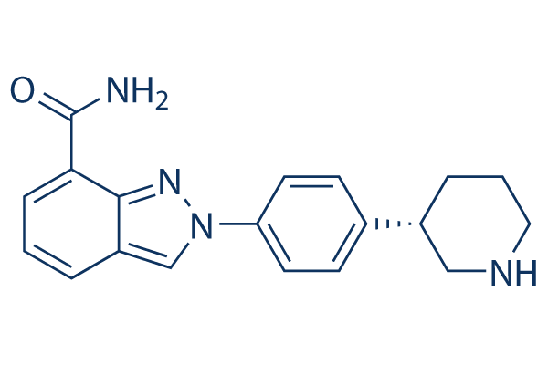
Chemische structuur
Moleculair gewicht: 320.39
Kwaliteitscontrole
| Gerelateerde doelwitten | HDAC ATM/ATR DNA-PK WRN DNA/RNA Synthesis Topoisomerase PPAR Sirtuin Casein Kinase eIF |
|---|---|
| Overige PARP Inhibitoren | XAV-939 AZD5305 (Saruparib) Veliparib (ABT-888) PJ34 HCl AG-14361 Iniparib (BSI-201) G007-LK Pamiparib UPF 1069 A-966492 |
Celkweek, behandeling & werkconcentratie
| Cellijnen | Assaytype | Concentratie | Incubatietijd | Formulering | Activiteitsbeschrijving | PMID |
|---|---|---|---|---|---|---|
| HeLa cells | Function assay | Inhibition of PARP in hydrogen peroxide-induced human HeLa cells assessed as inhibition DNA-damage-induced PARylation, EC50=0.004 μM | 19873981 | |||
| A549 cells | Cytotoxicity assay | 5-7 days | Cytotoxicity against human A549 cells transfected with BRCA2 shRNA assessed as inhibition of cell proliferation after 5 to 7 days by CellTiter-Blue assay, CC50=0.011 μM | 25761096 | ||
| MDA-MB-436 cells | Proliferation assay | 6 days | Antiproliferative activity against human MDA-MB-436 cells expressing BRCA1 5396 + 1G>A mutant after 6 days by cell titer-blue assay, CC50=18 nM | 19873981 | ||
| SUM1315MO2 cells | Cytotoxicity assay | 12 days | Cytotoxicity against human SUM1315MO2 cells carrying BRCA1 mutant assessed as inhibition of cell proliferation after 12 days by CellTiter-Blue assay, CC50=0.02 μM | 25761096 | ||
| DoTc2-4510 cells | Cytotoxicity assay | 5-7 days | Cytotoxicity against human DoTc2-4510 cells carrying BRCA2 mutant assessed as inhibition of cell proliferation after 5 to 7 days by CellTiter-Blue assay, CC50=0.023 μM | 25761096 | ||
| SUM149PT cells | Cytotoxicity assay | 5-7 days | Cytotoxicity against human SUM149PT cells carrying BRCA1 mutant assessed as inhibition of cell proliferation after 5 to 7 days by CellTiter-Blue assay, CC50=0.024 μM | 25761096 | ||
| UWB1.289 cells | Cytotoxicity assay | 5-7 days | Cytotoxicity against human UWB1.289 cells carrying BRCA1 mutant assessed as inhibition of cell proliferation after 5 to 7 days by CellTiter-Blue assay, CC50=0.056 μM | 25761096 | ||
| Capan1 cells | Cytotoxicity assay | Cytotoxicity against BRCA2-deficient human Capan1 cells, CC50=0.09 μM | 25761096 | |||
| Jurkat cells | Function assay | Inhibition of PARP1 in human Jurkat cells assessed as reduction of cell viability after 96 hrs by MTS assay in presence of 100 uM of temozolomide, EC50=0.2 μM | 23850199 | |||
| BT20 cells | Cytotoxicity assay | 5-7 days | Cytotoxicity against human BT20 cells assessed as inhibition of cell proliferation after 5 to 7 days by CellTiter-Blue assay, CC50=2.2 μM | 25761096 | ||
| Antiproliferative assay | HeLa | 7 days | Antiproliferative activity against BRCA1 deficient human HeLa cells after 7 days by cell titer-blue assay, CC50 = 0.033 μM. | 19873981 | ||
| Function assay | HeLa | Inhibition of PARP in hydrogen peroxide-induced human HeLa cells assessed as inhibition DNA-damage-induced PARylation, EC90 = 0.045 μM. | 19873981 | |||
| Antiproliferative assay | Capan1 | 13 days | Antiproliferative activity against human Capan1 cells expressing BRCA2 6174delT mutation and loss of wild-type allele after 13 days by cell titer-blue assay, CC50 = 0.09 μM. | 19873981 | ||
| Antiproliferative assay | HeLa | 7 days | Antiproliferative activity against human HeLa cells expressing wild type BRCA1 after 7 days by cell titer-blue assay, CC50 = 0.86 μM. | 19873981 | ||
| Function assay | Jurkat | 96 hrs | Inhibition of PARP1 in human Jurkat cells assessed as reduction of cell viability after 96 hrs by MTS assay, EC50 = 31 μM. | 23850199 | ||
| Function assay | CAPAN-1 | Inhibition of PARP in BRCA2-deficient human CAPAN-1 cells assessed as inhibition of hydrogen peroxide-induced PARylation by cell-based assay, IC50 = 0.0035 μM. | 25761096 | |||
| Function assay | HeLa | Inhibition of PARP in human HeLa cells assessed as inhibition of hydrogen peroxide-induced PARylation by cell-based assay, EC50 = 0.004 μM. | 25761096 | |||
| Function assay | A2780 | Inhibition of PARP in human A2780 cells assessed as inhibition of hydrogen peroxide-induced PARylation by cell-based assay, IC50 = 0.004 μM. | 25761096 | |||
| Cytotoxicity assay | MDA-MB-436 | Cytotoxicity against human MDA-MB-436 cells carrying BRCA1 mutant assessed as inhibition of cell proliferation, CC50 = 0.018 μM. | 25761096 | |||
| Cytotoxicity assay | HeLa | 5 to 7 days | Cytotoxicity against human HeLa cells transfected with BRCA1 shRNA assessed as reduction of cell viability after 5 to 7 days by CellTiter-Blue assay, CC50 = 0.034 μM. | 25761096 | ||
| Function assay | HeLa | Inhibition of PARP in human HeLa cells assessed as inhibition of hydrogen peroxide-induced PARylation by cell-based assay, IC90 = 0.046 μM. | 25761096 | |||
| Function assay | CAPAN-1 | Inhibition of PARP in BRCA2-deficient human CAPAN-1 cells assessed as inhibition of hydrogen peroxide-induced PARylation by cell-based assay, IC90 = 0.05 μM. | 25761096 | |||
| Function assay | A2780 | Inhibition of PARP in human A2780 cells assessed as inhibition of hydrogen peroxide-induced PARylation by cell-based assay, IC90 = 0.052 μM. | 25761096 | |||
| Cytotoxicity assay | HeLa | 5 to 7 days | Cytotoxicity against wild type human HeLa cells assessed as inhibition of cell proliferation after 5 to 7 days by CellTiter-Blue assay, CC50 = 0.852 μM. | 25761096 | ||
| Cytotoxicity assay | UWB1.289 | 5 to 7 days | Cytotoxicity against human UWB1.289 cells expressing BRCA1 assessed as inhibition of cell proliferation after 5 to 7 days by CellTiter-Blue assay, CC50 = 0.975 μM. | 25761096 | ||
| Cytotoxicity assay | A549 | 5 to 7 days | Cytotoxicity against wild type human A549 cells assessed as inhibition of cell proliferation after 5 to 7 days by CellTiter-Blue assay, CC50 = 1.76 μM. | 25761096 | ||
| Cytotoxicity assay | Capan1 | Cytotoxicity against BRCA2-deficient human Capan1 cells, EC50 = 0.65 μM. | 26652717 | |||
| Antitumor assay | MDA-MB-436 | 50 mg/kg | 33 days | Antitumor activity against human MDA-MB-436 cells expressing BRCA1 5396 + 1G>A mutant xenografted in CD1 mouse assessed as tumor regression at 50 mg/kg, po bid for 33 days | 19873981 | |
| Antitumor assay | MDA-MB-436 | 100 mg/kg | 33 days | Antitumor activity against human MDA-MB-436 cells expressing BRCA1 5396 + 1G>A mutant xenografted in CD1 mouse assessed as tumor regression at 100 mg/kg, po qd for 33 days | 19873981 | |
| Antitumor assay | MDA-MB-436 | 80 mg/kg | 1 to 2 weeks | Antitumor activity against human MDA-MB-436 cells harboring BRCA1 mutant xenografted in immunocompromised mouse assessed as tumor growth inhibition at 80 mg/kg, po qd for 1 to 2 weeks | 25761096 | |
| Antitumor assay | MDA-MB-436 | 80 mg/kg | 4 weeks | Antitumor activity against human MDA-MB-436 cells harboring BRCA1 mutant xenografted in immunocompromised mouse assessed as complete and sustained tumor regression at 80 mg/kg, po qd for 4 weeks | 25761096 | |
| Antitumor assay | MDA-MB-436 | 50 mg/kg | Antitumor activity against human MDA-MB-436 cells harboring BRCA1 mutant xenografted in immunocompromised mouse assessed as tumor growth inhibition at 50 mg/kg, po administered daily | 25761096 | ||
| Antitumor assay | MDA-MB-436 | 80 mg/kg | 3 weeks | Antitumor activity against human MDA-MB-436 cells harboring BRCA1 mutant xenografted in immunocompromised mouse assessed as tumor shrinkage at 80 mg/kg, po qd for 3 weeks | 25761096 | |
| qHTS assay | TC32 | qHTS of pediatric cancer cell lines to identify multiple opportunities for drug repurposing: Primary screen for TC32 cells | 29435139 | |||
| qHTS assay | A673 | qHTS of pediatric cancer cell lines to identify multiple opportunities for drug repurposing: Primary screen for A673 cells | 29435139 | |||
| qHTS assay | NB1643 | qHTS of pediatric cancer cell lines to identify multiple opportunities for drug repurposing: Primary screen for NB1643 cells | 29435139 | |||
| qHTS assay | A673 | qHTS of pediatric cancer cell lines to identify multiple opportunities for drug repurposing: Confirmatory screen for A673 cells) | 29435139 | |||
| qHTS assay | BT-37 | qHTS of pediatric cancer cell lines to identify multiple opportunities for drug repurposing: Confirmatory screen for BT-37 cells | 29435139 | |||
| qHTS assay | SK-N-MC | qHTS of pediatric cancer cell lines to identify multiple opportunities for drug repurposing: Primary screen for SK-N-MC cells | 29435139 | |||
| qHTS assay | NB-EBc1 | qHTS of pediatric cancer cell lines to identify multiple opportunities for drug repurposing: Primary screen for NB-EBc1 cells | 29435139 | |||
| qHTS assay | LAN-5 | qHTS of pediatric cancer cell lines to identify multiple opportunities for drug repurposing: Primary screen for LAN-5 cells | 29435139 | |||
| qHTS assay | SK-N-MC | qHTS of pediatric cancer cell lines to identify multiple opportunities for drug repurposing: Confirmatory screen for SK-N-MC cells | 29435139 | |||
| qHTS assay | TC32 | qHTS of pediatric cancer cell lines to identify multiple opportunities for drug repurposing: Confirmatory screen for TC32 cells | 29435139 | |||
| Klik om meer experimentele gegevens over de cellijn te bekijken | ||||||
Chemische informatie, Opslag en Stabiliteit
| Moleculair gewicht | 320.39 | Formule | C19H20N4O |
Opslag (Vanaf de ontvangstdatum) | |
|---|---|---|---|---|---|
| CAS-nr. | 1038915-60-4 | SDF downloaden | Opslag van stamoplossingen |
|
|
Oplosbaarheid
|
In vitro |
DMSO
: 64 mg/mL
(199.75 mM)
Ethanol : 64 mg/mL Water : Insoluble |
Molariteitscalculator
|
In vivo |
|||||
In vivo Formuleringscalculator (Heldere oplossing)
Stap 1: Voer de onderstaande informatie in (Aanbevolen: Een extra dier voor het geval van verlies tijdens het experiment)
Stap 2: Voer de in vivo formulering in (Dit is alleen de calculator, geen formulering. Neem eerst contact met ons op als er geen in vivo formulering is in het gedeelte Oplosbaarheid.)
Berekeningsresultaten:
Werkconcentratie: mg/ml;
Methode voor het bereiden van DMSO-mastervloeistof: mg geneesmiddel vooraf opgelost in μL DMSO ( Concentratie mastervloeistof mg/mL, Neem eerst contact met ons op als de concentratie de DMSO-oplosbaarheid van de partij geneesmiddel overschrijdt. )
Methode voor het bereiden van in vivo formulering: Neem μL DMSO mastervloeistof, voeg vervolgens toeμL PEG300, mengen en helder maken, voeg vervolgens toeμL Tween 80, mengen en helder maken, voeg vervolgens toe μL ddH2O, mengen en helder maken.
Methode voor het bereiden van in vivo formulering: Neem μL DMSO mastervloeistof, voeg vervolgens toe μL Maïsolie, mengen en helder maken.
Opmerking: 1. Zorg ervoor dat de vloeistof helder is voordat u het volgende oplosmiddel toevoegt.
2. Zorg ervoor dat u het/de oplosmiddel(en) in de juiste volgorde toevoegt. U moet ervoor zorgen dat de verkregen oplossing, bij de vorige toevoeging, een heldere oplossing is voordat u verdergaat met het toevoegen van het volgende oplosmiddel. Fysische methoden zoals vortexen, echografie of een warmwaterbad kunnen worden gebruikt om het oplossen te bevorderen.
Werkingsmechanisme
| Targets/IC50/Ki |
PARP2
(Cell-free assay) 2.1 nM
PARP1
(Cell-free assay) 3.8 nM
|
|---|---|
| In vitro |
In een hele celassay remde Niraparib (MK-4827) de PARP-activiteit met EC50 = 4 nM en remde de proliferatie van kankercellen met mutante BRCA-1 en BRCA-2 met CC50 in het bereik van 10-100 nM. Het bleek een potente en selectieve PARP-1- en PARP-2-remmer te zijn met respectievelijk IC50=3,8 en 2,1 nM. Bovendien vertoonde deze verbinding een ten minste 100-voudige selectiviteit ten opzichte van PARP-3, V-PARP en tankyrase-1, met respectievelijk IC50 = 1300, 330 en 570 nM. Naast het remmen van de groei van HeLa-cellen die BRCA-1 missen als gevolg van silencing door RNA-interferentie, is het in staat de proliferatie van kankercellijnen met natuurlijke BRCA-1- of BRCA-2-mutaties te remmen. In MDA-MB-436 menselijke borstklieradenocarcinoomcellen met BRCA-1-mutaties vertoonde het CC50 = 18 nM, terwijl in CAPAN-1 menselijke pancreasklieradenocarcinoomcellen, die BRCA-2-mutant zijn, het CC50 = 90 nM vertoonde. Daarentegen zijn normale menselijke prostaat- en mamma-epitheelcellen resistent tegen MK-4827, waarbij antiproliferatieve effecten in het micromolaire bereik worden waargenomen, wat de zeer hoge selectieve cytotoxiciteit van deze PARP-remmers in BRCA-1- en -2-mutante kankercellen aantoont in vergelijking met het omringende weefsel.
|
| In vivo |
Niraparib (MK-4827), een nieuwe, oraal biologisch beschikbare PARP-1- en PARP-2-remmer, versterkte sterk het effect van straling op een verscheidenheid aan menselijke tumorxenografts, zowel p53 wild type als p53 mutant. Het werd goed verdragen in vivo en toonde werkzaamheid als een enkelvoudig middel in een xenograftmodel van BRCA-1-deficiënte kanker.
|
Referenties |
|
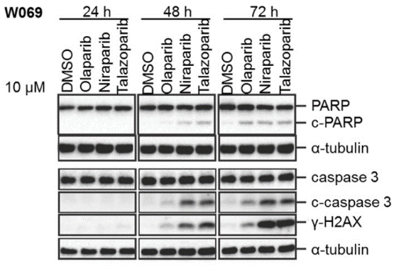
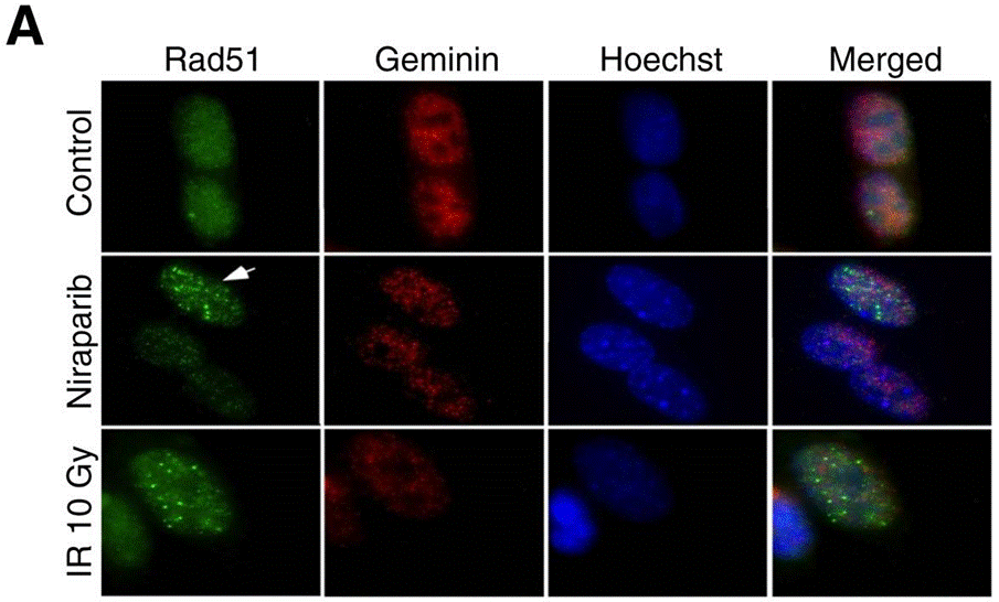
Toepassingen
| Methoden | Biomarkers | Afbeeldingen | PMID |
|---|---|---|---|
| Western blot | c-PARP /c-caspase 3 / γ-H2AX |
|
29158830 |
| Immunofluorescence | Rad51 / Geminin |
|
27614696 |
Informatie klinische proef
(gegevens van https://clinicaltrials.gov, bijgewerkt op 2024-05-22)
| NCT-nummer | Rekrutering | Aandoeningen | Sponsor/Medewerkers | Startdatum | Fasen |
|---|---|---|---|---|---|
| NCT05289648 | Not yet recruiting | Endometrial Cancer|Serous Adenocarcinoma|Uterine Neoplasm |
Sir Mortimer B. Davis - Jewish General Hospital |
May 1 2024 | Early Phase 1 |
| NCT06077877 | Recruiting | Neoplasms |
GlaxoSmithKline |
October 24 2023 | Phase 1|Phase 2 |
| NCT05666349 | Withdrawn | Recurrent Glioblastoma |
University College London|GlaxoSmithKline |
October 13 2023 | Phase 1 |
Technische ondersteuning
Tel: +1-832-582-8158 Ext:3
Als u nog andere vragen heeft, kunt u een bericht achterlaten.
Veelgestelde vragen
Vraag 1:
How to reconstitute it for in vivo studies?
Antwoord:
It can be orally administered using the formulation 1% CMC-Na (suspension).
Producten zijn uitsluitend voor onderzoeksdoeleinden. Niet voor menselijk gebruik. Wij verkopen niet aan patiënten.
©Copyright 2013 Selleck Chemicals. Alle rechten voorbehouden.