|
Come citare 1. Per la citazione nel testo (Materiali e Metodi): 2. Per la tabella delle risorse chiave: |
||
|
Numero verde: (877) 796-6397 -- Solo USA e Canada -- |
Fax: +1-832-582-8590 Ordini: +1-832-582-8158 |
Supporto tecnico: +1-832-582-8158 Ext:3 Si prega di fornire il numero dordine nelle-mail. Ci sforziamo di rispondere a tutte le richieste via e-mail entro un giorno lavorativo. |
Descrizione biologica
| Specificità | ZO1 tight junction protein Antibody [A24G22] rileva i livelli endogeni della proteina totale ZO1 tight junction protein. |
|---|---|
| Contesto | ZO-1 (Zonula Occludens-1) è una fosfoproteina di membrana periferica appartenente alla famiglia delle guanilato chinasi associate alla membrana (MAGUK) e funge da molecola di scaffolding critica nelle giunzioni strette. ZO-1 contiene tre domini PDZ che facilitano le interazioni proteina-proteina, un dominio SH3 che lega varie proteine di segnalazione e un dominio guanilato chinasi (GuK) collegato da regioni cerniera flessibili; la sua unica regione C-terminale si lega direttamente al citoscheletro di actina, ancorando il complesso della giunzione stretta per mantenere l'integrità cellulare. ZO-1 organizza le proteine transmembrana delle giunzioni strette come occludina e claudine, collegandole a partner citosolici come ZO-2 e ZO-3, svolgendo così un ruolo essenziale nell'assemblaggio e nel mantenimento della barriera delle giunzioni strette che controlla la permeabilità paracellulare. ZO-1 agisce anche come hub di segnalazione che regola la crescita cellulare, la migrazione, l'angiogenesi e l'infiammazione e aiuta a mantenere la polarità delle cellule epiteliali preservando il confine tra i domini di membrana apicali e basolaterali. ZO-1 è vitale per l'omeostasi tissutale in barriere come l'epitelio intestinale e la barriera emato-encefalica, con la sua downregulation associata a malattie come la malattia infiammatoria intestinale (IBD) e la progressione del cancro, dove le giunzioni strette compromesse contribuiscono alla permeabilità patologica e alla tumorigenesi. |
Informazioni sullutilizzo
| Applicazione | IHC, IF, FCM | Diluizione |
|
||||||
|---|---|---|---|---|---|---|---|---|---|
| Reattività | Rat, Human, Mouse | ||||||||
| Fonte | Rabbit Monoclonal Antibody | MW | |||||||
| Tampone di conservazione | PBS, pH 7.2+50% Glycerol+0.05% BSA+0.01% NaN3 | Conservazione (Dalla data di ricevimento) |
-20°C (avoid freeze-thaw cycles), 2 years | ||||||
| IHC |
Experimental Protocol:
Deparaffinization/Rehydration
1. Deparaffinize/hydrate sections:
2. Incubate sections in three washes of xylene for 5 min each.
3. Incubate sections in two washes of 100% ethanol for 10 min each.
4. Incubate sections in two washes of 95% ethanol for 10 min each.
5. Wash sections two times in dH2O for 5 min each.
6.Antigen retrieval: For Citrate: Heat slides in a microwave submersed in 1X citrate unmasking solution until boiling is initiated; continue with 10 min at a sub-boiling temperature (95°-98°C). Cool slides on bench top for 30 min.
Staining
1. Wash sections in dH2O three times for 5 min each.
2. Incubate sections in 3% hydrogen peroxide for 10 min.
3. Wash sections in dH2O two times for 5 min each.
4. Wash sections in wash buffer for 5 min.
5. Block each section with 100–400 µl of blocking solution for 1 hr at room temperature.
6. Remove blocking solution and add 100–400 µl primary antibody diluent in to each section. Incubate overnight at 4°C.
7. Remove antibody solution and wash sections with wash buffer three times for 5 min each.
8. Cover section with 1–3 drops HRPas needed. Incubate in a humidified chamber for 30 min at room temperature.
9. Wash sections three times with wash buffer for 5 min each.
10. Add DAB Chromogen Concentrate to DAB Diluent and mix well before use.
11. Apply 100–400 µl DAB to each section and monitor closely. 1–10 min generally provides an acceptable staining intensity.
12. Immerse slides in dH2O.
13. If desired, counterstain sections with hematoxylin.
14. Wash sections in dH2O two times for 5 min each.
15. Dehydrate sections: Incubate sections in 95% ethanol two times for 10 sec each; Repeat in 100% ethanol, incubating sections two times for 10 sec each; Repeat in xylene, incubating sections two times for 10 sec each.
16. Mount sections with coverslips and mounting medium.
|
| IF |
Experimental Protocol:
Sample Preparation
1. Adherent Cells: Place a clean, sterile coverslip in a culture dish. Once the cells grow to near confluence as a monolayer, remove the coverslip for further use.
2. Suspension Cells: Seed the cells onto a clean, sterile slide coated with poly-L-lysine.
3. Frozen Sections: Allow the slide to thaw at room temperature. Wash it with pure water or PBS for 2 times, 3 minutes each time.
4. Paraffin Sections: Deparaffinization and rehydration. Wash the slide with pure water or PBS for 3 times, 3 minutes each time. Then perform antigen retrieval.
Fixation
1. Fix the cell coverslips/spots or tissue sections at room temperature using a fixative such as 4% paraformaldehyde (4% PFA) for 10-15 minutes.
2. Wash the sample with PBS for 3 times, 3 minutes each time.
Permeabilization
1.Add a detergent such as 0.1–0.3% Triton X-100 to the sample and incubate at room temperature for 10–20 minutes.
(Note: This step is only required for intracellular antigens. For antigens expressed on the cell membrane, this step is unnecessary.)
Wash the sample with PBS for 3 times, 3 minutes each time.
Blocking
Add blocking solution and incubate at room temperature for at least 1 hour. (Common blocking solutions include: serum from the same source as the secondary antibody, BSA, or goat serum.)
Note: Ensure the sample remains moist during and after the blocking step to prevent drying, which can lead to high background.
Immunofluorescence Staining (Day 1)
1. Remove the blocking solution and add the diluted primary antibody.
2. Incubate the sample in a humidified chamber at 4°C overnight.
Immunofluorescence Staining (Day 2)
1. Remove the primary antibody and wash with PBST for 3 times, 5 minutes each time.
2. Add the diluted fluorescent secondary antibody and incubate in the dark at 4°C for 1–2 hours.
3. Remove the secondary antibody and wash with PBST for 3 times, 5 minutes each time.
4. Add diluted DAPI and incubate at room temperature in the dark for 5–10 minutes.
5. Wash with PBST for 3 times, 5 minutes each time.
Mounting
1. Mount the sample with an anti-fade mounting medium.
2. Allow the slide to dry at room temperature overnight in the dark.
3. Store the slide in a slide storage box at 4°C, protected from light.
|
Riferimenti
|
Dati di applicazione
IF
Validato da Selleck
-
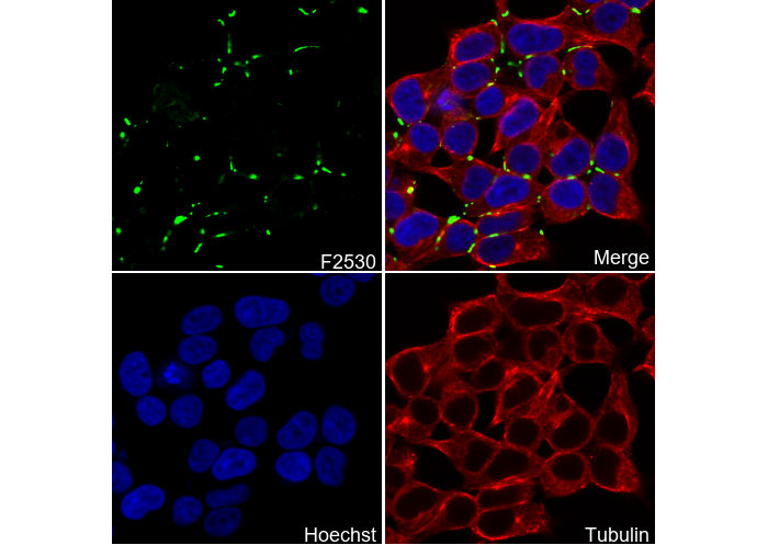 Immunofluorescent analysis of 293T cells using F2530 (green, 1:100), Hoechst (blue) and tubulin (Red).