|
Come citare 1. Per la citazione nel testo (Materiali e Metodi): 2. Per la tabella delle risorse chiave: |
||
|
Numero verde: (877) 796-6397 -- Solo USA e Canada -- |
Fax: +1-832-582-8590 Ordini: +1-832-582-8158 |
Supporto tecnico: +1-832-582-8158 Ext:3 Si prega di fornire il numero dordine nelle-mail. Ci sforziamo di rispondere a tutte le richieste via e-mail entro un giorno lavorativo. |
Descrizione biologica
| Specificità | Wnt3 and Wnt3a Antibody [J20B9] rileva i livelli endogeni della proteina Wnt3 e Wnt3a totale. |
|---|---|
| Contesto | Wnt3 e Wnt3a sono glicoproteine secrete ricche di cisteina, strettamente correlate, della famiglia Wnt che agiscono come potenti ligandi per la via canonica Wnt/beta-catenina, controllando il patterning embrionale, il mantenimento delle cellule staminali e l'omeostasi tissutale. Ogni proteina è composta da ~350-400 amminoacidi e adotta la caratteristica architettura Wnt a “pollice-indice” con domini N-terminale e C-terminale stabilizzati da molteplici legami disolfuro e modificati da N-glicosilazione e da una cruciale O-palmitoleoilazione su una serina conservata (Ser212 in Wnt3, Ser209 in Wnt3a), che è necessaria per il legame al trasportatore Wntless per la secrezione e per l'engagement ad alta affinità dei recettori Frizzled. Wnt3 e Wnt3a si legano ai complessi recettoriali Frizzled/LRP5-6 sulla superficie cellulare per reclutare e attivare Dishevelled, inibire il complesso di distruzione Axin/APC/CK1/GSK-3β, stabilizzare la β-catenina citosolica e guidarne l'accumulo nucleare dove si associa a TCF/LEF per indurre geni target come cc-MYC, CCND1 (ciclina D1), AXIN2 e HOXB4 che promuovono la proliferazione, la sopravvivenza e la staminalità; Wnt3 e Wnt3a sono quindi essenziali per la formazione dell'asse, lo sviluppo degli arti e neurale, e la regolazione delle cellule staminali ematopoietiche, neurali e intestinali, con effetti parzialmente sovrapposti ma specifici per il contesto (ad esempio, Wnt3a che sostiene la proliferazione dei precursori neurali, Wnt3 che orienta più fortemente verso la neurogenesi in alcuni domini del midollo spinale). Mutazioni con perdita di funzione in WNT3 causano tetra-amelia autosomica recessiva con gravi malformazioni degli arti e craniofacciali, mentre l'aberrante up-regolazione della segnalazione della β-catenina mediata da Wnt3/Wnt3a contribuisce all'oncogenesi nei tumori colorettali, della mammella, del polmone e in altri tumori, promuovendo un'entrata incontrollata nel ciclo cellulare, l'EMT e la resistenza all'apoptosi, e una segnalazione Wnt3a disregolata è stata anche implicata nei disturbi scheletrici e neurodegenerativi. |
Informazioni sullutilizzo
| Applicazione | WB | Diluizione |
|
||
|---|---|---|---|---|---|
| Reattività | Human | ||||
| Fonte | Rabbit Monoclonal Antibody | MW | 39 kDa | ||
| Tampone di conservazione | PBS, pH 7.2+50% Glycerol+0.05% BSA+0.01% NaN3 | Conservazione (Dalla data di ricevimento) |
-20°C (avoid freeze-thaw cycles), 2 years | ||
| WB |
Experimental Protocol:
Sample preparation
1. Tissue: Lyse the tissue sample by adding an appropriate volume of ice-cold RIPA/NP-40 Lysis Buffer (containing Protease Inhibitor Cocktail),and homogenize the tissue at a low temperature. 2. Adherent cell: Aspirate the culture medium and wash the cells with ice-cold PBS twice. Lyse the cells by adding an appropriate volume of RIPA/NP-40 Lysis Buffer (containing Protease Inhibitor Cocktail) and put the sample on ice for 5 min. 3. Suspension cell: Transfer the culture medium to a pre-cooled centrifuge tube. Centrifuge and aspirate the supernatant. Wash the cells with ice-cold PBS twice. Lyse the cells by adding an appropriate volume of RIPA/NP-40 Lysis Buffer (containing Protease Inhibitor Cocktail) and put the sample on ice for 5 min. 4. Place the lysate into a pre-cooled microcentrifuge tube. Centrifuge at 4°C for 15 min. Collect the supernatant;
5. Remove a small volume of lysate to determine the protein concentration;
6. Combine the lysate with protein loading buffer. Boil 20 µL sample under 95-100°C for 5 min. Centrifuge for 5 min after cool down on ice.
Electrophoretic separation
1. According to the concentration of extracted protein, load appropriate amount of protein sample and marker onto SDS-PAGE gels for electrophoresis. Recommended separating gel (lower gel) concentration: 10%. Reference Table for Selecting SDS-PAGE Separation Gel Concentrations 2. Power up 80V for 30 minutes. Then the power supply is adjusted (110 V~150 V), the Marker is observed, and the electrophoresis can be stopped when the indicator band of the predyed protein Marker where the protein is located is properly separated. (Note that the current should not be too large when electrophoresis, too large current (more than 150 mA) will cause the temperature to rise, affecting the result of running glue. If high currents cannot be avoided, an ice bath can be used to cool the bath.)
Transfer membrane
1. Take out the converter, soak the clip and consumables in the pre-cooled converter;
2. Activate PVDF membrane with methanol for 1 min and rinse with transfer buffer;
3. Install it in the order of "black edge of clip - sponge - filter paper - filter paper - glue -PVDF membrane - filter paper - filter paper - sponge - white edge of clip"; 4. The protein was electrotransferred to PVDF membrane. ( 0.45 µm PVDF membrane is recommended ) Reference Table for Selecting PVDF Membrane Pore Size Specifications Recommended conditions for wet transfer: 200 mA, 60 min. ( Note that the transfer conditions can be adjusted according to the protein size. For high-molecular-weight proteins, a higher current and longer transfer time are recommended. However, ensure that the transfer tank remains at a low temperature to prevent gel melting.)
Block
1. After electrotransfer, wash the film with TBST at room temperature for 5 minutes;
2. Incubate the film in the blocking solution for 1 hour at room temperature;
3. Wash the film with TBST for 3 times, 5 minutes each time.
Antibody incubation
1. Use 5% skim milk powder to prepare the primary antibody working liquid (recommended dilution ratio for primary antibody 1:10000), gently shake and incubate with the film at 4°C overnight; 2. Wash the film with TBST 3 times, 5 minutes each time;
3. Add the secondary antibody to the blocking solution and incubate with the film gently at room temperature for 1 hour;
4. After incubation, wash the film with TBST 3 times for 5 minutes each time.
Antibody staining
1. Add the prepared ECL luminescent substrate (or select other color developing substrate according to the second antibody) and mix evenly;
2. Incubate with the film for 1 minute, remove excess substrate (keep the film moist), wrap with plastic film, and expose in the imaging system.
|
Riferimenti
|
Dati di applicazione
WB
Validato da Selleck
-
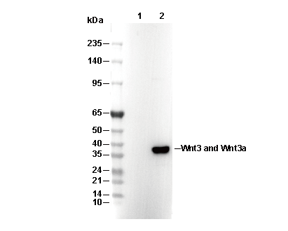 Lane 1: CHO, Lane 2: CHO(Human Wnt3a transfected)