|
Come citare 1. Per la citazione nel testo (Materiali e Metodi): 2. Per la tabella delle risorse chiave: |
||
|
Numero verde: (877) 796-6397 -- Solo USA e Canada -- |
Fax: +1-832-582-8590 Ordini: +1-832-582-8158 |
Supporto tecnico: +1-832-582-8158 Ext:3 Si prega di fornire il numero dordine nelle-mail. Ci sforziamo di rispondere a tutte le richieste via e-mail entro un giorno lavorativo. |
Descrizione biologica
| Specificità | TSH Receptor/TSH-R Antibody [L10M22] rileva i livelli endogeni della proteina TSH Receptor/TSH-R totale. |
|---|---|
| Contesto | Il recettore dell'ormone tireostimolante (TSHR) è un regolatore chiave del metabolismo dell'ormone tiroideo e funge da controllore primario della funzione e della crescita delle cellule tiroidee. Appartiene alla famiglia dei recettori accoppiati a proteine G con sette domini transmembrana ed è posizionato sulla membrana basolaterale delle cellule follicolari tiroidee. La proteina TSHR è composta da 764 aminoacidi, ha un peso molecolare di circa 87 kDa e media i suoi effetti attraverso l'interazione con più sottotipi di proteine G, in particolare Gαs e Gαq. Dopo l'attivazione da parte del TSH, il recettore avvia la segnalazione intracellulare attraverso queste proteine G, modulando così l'attività delle molecole effettrici a valle. Il percorso Gαs stimola la cascata del monofosfato di adenosina ciclico (cAMP), mentre il percorso Gαq attiva la cascata della fosfolipasi C (PLC). A concentrazioni elevate di TSH, il cAMP si lega alla protein chinasi A (PKA), che fosforila vari effettori bersaglio, migliorandone l'attività catalitica. In parallelo, l'attivazione della PLC genera inositolo 1,4,5-trifosfato (IP₃) e diacilglicerolo (DAG), amplificando ulteriormente le risposte cellulari. L'espressione di TSHR è regolata positivamente dai livelli fisiologici di TSH ma viene downregolata a concentrazioni di TSH persistentemente elevate. La sovrastimolazione cronica del TSHR, in particolare attraverso il percorso del cAMP, può portare a un'eccessiva secrezione di ormoni tiroidei, iperplasia follicolare tiroidea e ipertiroidismo clinico. Le mutazioni nel gene TSHR possono influenzare la struttura proteica del recettore o le sue modifiche post-traduzionali, alterando così la funzione del recettore. Sebbene TSHR non avvii direttamente la cancerogenesi, può contribuire significativamente alla crescita tumorale quando gli oncogeni sono già attivati. |
Informazioni sullutilizzo
| Applicazione | IHC | Diluizione |
|
||
|---|---|---|---|---|---|
| Reattività | Human | ||||
| Fonte | Rabbit Monoclonal Antibody | MW | |||
| Tampone di conservazione | PBS, pH 7.2+50% Glycerol+0.05% BSA+0.01% NaN3 | Conservazione (Dalla data di ricevimento) |
-20°C (avoid freeze-thaw cycles), 2 years | ||
| IHC |
Experimental Protocol:
Deparaffinization/Rehydration
1. Deparaffinize/hydrate sections:
2. Incubate sections in three washes of xylene for 5 min each.
3. Incubate sections in two washes of 100% ethanol for 10 min each.
4. Incubate sections in two washes of 95% ethanol for 10 min each.
5. Wash sections two times in dH2O for 5 min each.
6.Antigen retrieval: For Citrate: Heat slides in a microwave submersed in 1X citrate unmasking solution until boiling is initiated; continue with 10 min at a sub-boiling temperature (95°-98°C). Cool slides on bench top for 30 min.
Staining
1. Wash sections in dH2O three times for 5 min each.
2. Incubate sections in 3% hydrogen peroxide for 10 min.
3. Wash sections in dH2O two times for 5 min each.
4. Wash sections in wash buffer for 5 min.
5. Block each section with 100–400 µl of blocking solution for 1 hr at room temperature.
6. Remove blocking solution and add 100–400 µl primary antibody diluent in to each section. Incubate overnight at 4°C.
7. Remove antibody solution and wash sections with wash buffer three times for 5 min each.
8. Cover section with 1–3 drops HRPas needed. Incubate in a humidified chamber for 30 min at room temperature.
9. Wash sections three times with wash buffer for 5 min each.
10. Add DAB Chromogen Concentrate to DAB Diluent and mix well before use.
11. Apply 100–400 µl DAB to each section and monitor closely. 1–10 min generally provides an acceptable staining intensity.
12. Immerse slides in dH2O.
13. If desired, counterstain sections with hematoxylin.
14. Wash sections in dH2O two times for 5 min each.
15. Dehydrate sections: Incubate sections in 95% ethanol two times for 10 sec each; Repeat in 100% ethanol, incubating sections two times for 10 sec each; Repeat in xylene, incubating sections two times for 10 sec each.
16. Mount sections with coverslips and mounting medium.
|
Riferimenti
|
Dati di applicazione
IHC
Validato da Selleck
-
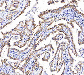 Immunohistochemical analysis of formalin fixed paraffin embedded human thyroid gland tissue with F3696 at 1:1000 dilution.