|
Come citare 1. Per la citazione nel testo (Materiali e Metodi): 2. Per la tabella delle risorse chiave: |
||
|
Numero verde: (877) 796-6397 -- Solo USA e Canada -- |
Fax: +1-832-582-8590 Ordini: +1-832-582-8158 |
Supporto tecnico: +1-832-582-8158 Ext:3 Si prega di fornire il numero dordine nelle-mail. Ci sforziamo di rispondere a tutte le richieste via e-mail entro un giorno lavorativo. |
Descrizione biologica
| Specificità | TRAF6 Antibody [B8B10] rileva i livelli endogeni della proteina TRAF6 totale. Questo anticorpo non dovrebbe reagire in modo crociato con altri membri della famiglia TRAF. |
|---|---|
| Contesto | I fattori associati al recettore del fattore di necrosi tumorale (TRAF) costituiscono una famiglia di proteine adattatrici citoplasmatiche che mediano diversi eventi di segnalazione intracellulare. Nei mammiferi, sono stati identificati sette membri della famiglia TRAF, inclusi sei membri classici (TRAF1-TRAF6) e un membro non classico (TRAF7). I TRAF classici condividono un dominio TRAF carboxy-terminale conservato, che è assente nel TRAF7 non classico. Funzionalmente, i TRAF partecipano alla trasduzione del segnale della superfamiglia del fattore di necrosi tumorale (TNFSF) così come ai membri della superfamiglia del recettore Toll-like/IL-1 (TLR/ILR), regolando così le cascate di segnalazione a valle come la via della chinasi proteica attivata da mitogeni (MAPK). Oltre alla segnalazione del recettore, i TRAF svolgono ruoli essenziali nella proliferazione, differenziazione, apoptosi e sopravvivenza cellulare, modulando anche le risposte immunitarie e infiammatorie. La disregolazione delle proteine TRAF contribuisce all'oncogenesi, con membri distinti che esercitano funzioni oncogeniche o di soppressione tumorale. TRAF1, TRAF2, TRAF4, TRAF5 e TRAF6 sono stati implicati nella carcinogenesi, mentre TRAF3 funziona prevalentemente come soppressore tumorale. Tra la famiglia, TRAF6 si distingue per la sua unica specificità di legame al recettore, consentendogli di interagire non solo con i membri della superfamiglia dei recettori TNF, ma anche con le proteine della superfamiglia IL-1R/TLR. L'iperespressione di TRAF6 è stata riportata in diversi tipi di tumore, inclusi i carcinomi del colon, gastrici e mammari, così come il melanoma. Funzionalmente, TRAF6 promuove l'inizio e la progressione del tumore modulando l'apoptosi, la proliferazione, la sopravvivenza e l'invasione. Meccanicamente, TRAF6 attiva diverse vie di segnalazione, con la cascata del recettore Toll-like 4 (TLR4) che è particolarmente significativa. Attraverso percorsi sia MyD88-dipendenti che MyD88-indipendenti, TRAF6 amplifica la segnalazione infiammatoria e di sopravvivenza. Inoltre, TRAF6 migliora l'attivazione della via PI3K-AKT facilitando l'ubiquitinazione di PI3K, aumentando così la fosforilazione di AKT e guidando la crescita cellulare. Collettivamente, questi risultati evidenziano TRAF6 come un adattatore critico che integra la segnalazione del recettore con i processi oncogenici. |
Informazioni sullutilizzo
| Applicazione | WB, IP | Diluizione |
|
||||
|---|---|---|---|---|---|---|---|
| Reattività | Human, Monkey | ||||||
| Fonte | Rabbit Monoclonal Antibody | MW | 60 kDa | ||||
| Tampone di conservazione | PBS, pH 7.2+50% Glycerol+0.05% BSA+0.01% NaN3 | Conservazione (Dalla data di ricevimento) |
-20°C (avoid freeze-thaw cycles), 2 years | ||||
| WB |
Experimental Protocol:
Sample preparation
1. Tissue: Lyse the tissue sample by adding an appropriate volume of ice-cold RIPA/NP-40 Lysis Buffer (containing Protease Inhibitor Cocktail),and homogenize the tissue at a low temperature. 2. Adherent cell: Aspirate the culture medium and wash the cells with ice-cold PBS twice. Lyse the cells by adding an appropriate volume of RIPA/NP-40 Lysis Buffer (containing Protease Inhibitor Cocktail) and put the sample on ice for 5 min. 3. Suspension cell: Transfer the culture medium to a pre-cooled centrifuge tube. Centrifuge and aspirate the supernatant. Wash the cells with ice-cold PBS twice. Lyse the cells by adding an appropriate volume of RIPA/NP-40 Lysis Buffer (containing Protease Inhibitor Cocktail) and put the sample on ice for 5 min. 4. Place the lysate into a pre-cooled microcentrifuge tube. Centrifuge at 4°C for 15 min. Collect the supernatant;
5. Remove a small volume of lysate to determine the protein concentration;
6. Combine the lysate with protein loading buffer. Boil 20 µL sample under 95-100°C for 5 min. Centrifuge for 5 min after cool down on ice.
Electrophoretic separation
1. According to the concentration of extracted protein, load appropriate amount of protein sample and marker onto SDS-PAGE gels for electrophoresis. Recommended separating gel (lower gel) concentration: 10%. Reference Table for Selecting SDS-PAGE Separation Gel Concentrations 2. Power up 80V for 30 minutes. Then the power supply is adjusted (110 V~150 V), the Marker is observed, and the electrophoresis can be stopped when the indicator band of the predyed protein Marker where the protein is located is properly separated. (Note that the current should not be too large when electrophoresis, too large current (more than 150 mA) will cause the temperature to rise, affecting the result of running glue. If high currents cannot be avoided, an ice bath can be used to cool the bath.)
Transfer membrane
1. Take out the converter, soak the clip and consumables in the pre-cooled converter;
2. Activate PVDF membrane with methanol for 1 min and rinse with transfer buffer;
3. Install it in the order of "black edge of clip - sponge - filter paper - filter paper - glue -PVDF membrane - filter paper - filter paper - sponge - white edge of clip"; 4. The protein was electrotransferred to PVDF membrane. ( 0.45 µm PVDF membrane is recommended ) Reference Table for Selecting PVDF Membrane Pore Size Specifications Recommended conditions for wet transfer: 200 mA, 120 min. ( Note that the transfer conditions can be adjusted according to the protein size. For high-molecular-weight proteins, a higher current and longer transfer time are recommended. However, ensure that the transfer tank remains at a low temperature to prevent gel melting.)
Block
1. After electrotransfer, wash the film with TBST at room temperature for 5 minutes;
2. Incubate the film in the blocking solution for 1 hour at room temperature;
3. Wash the film with TBST for 3 times, 5 minutes each time.
Antibody incubation
1. Use 5% skim milk powder to prepare the primary antibody working liquid (recommended dilution ratio for primary antibody 1:1000), gently shake and incubate with the film at 4°C overnight; 2. Wash the film with TBST 3 times, 5 minutes each time;
3. Add the secondary antibody to the blocking solution and incubate with the film gently at room temperature for 1 hour;
4. After incubation, wash the film with TBST 3 times for 5 minutes each time.
Antibody staining
1. Add the prepared ECL luminescent substrate (or select other color developing substrate according to the second antibody) and mix evenly;
2. Incubate with the film for 1 minute, remove excess substrate (keep the film moist), wrap with plastic film, and expose in the imaging system.
|
Riferimenti
|
Dati di applicazione
WB
Validato da Selleck
-
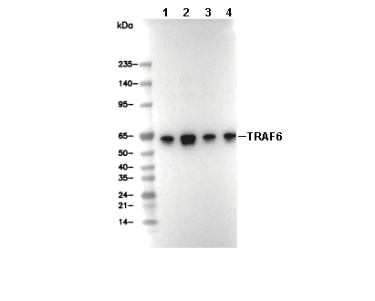 Lane 1: K562, Lane 2: Hela, Lane 3: 293T, Lane 4: COS-7