|
Come citare 1. Per la citazione nel testo (Materiali e Metodi): 2. Per la tabella delle risorse chiave: |
||
|
Numero verde: (877) 796-6397 -- Solo USA e Canada -- |
Fax: +1-832-582-8590 Ordini: +1-832-582-8158 |
Supporto tecnico: +1-832-582-8158 Ext:3 Si prega di fornire il numero dordine nelle-mail. Ci sforziamo di rispondere a tutte le richieste via e-mail entro un giorno lavorativo. |
Descrizione biologica
| Specificità | TGF β Receptor II Antibody [J15L22] rileva i livelli endogeni della proteina TGF-β Receptor II totale. |
|---|---|
| Contesto | Il TGF β Receptor II (recettore del fattore di crescita trasformante-β di tipo II, TβRII) è una glicoproteina transmembrana di tipo I e una serina/treonina chinasi costitutivamente attiva che avvia la segnalazione di TGF-β. Comprende un ectodominio extracellulare di legame al ligando con una piega tossinica a tre dita stabilizzata da sei legami disolfuro (quattro conservati tra i recettori di tipo II e due unici per TβRII), un singolo segmento transmembrana e un dominio chinasico intracellulare. L'ectodominio contiene 12 cisteine e presenta un primo “dito” esteso rispetto ai recettori correlati, consentendo interazioni specifiche con il ligando. Il TβRII è espresso in molti tessuti e si lega ai ligandi di TGF-β, reclutando e fosforilando il recettore di tipo I (TβRI) per attivare le vie dipendenti da SMAD e indipendenti da SMAD, inclusa la segnalazione della chinasi Src. Attraverso questi meccanismi, il TβRII regola lo sviluppo, l'omeostasi tissutale, le risposte immunitarie e i processi patologici come il cancro e la fibrosi. |
Informazioni sullutilizzo
| Applicazione | IHC, FCM | Diluizione |
|
||
|---|---|---|---|---|---|
| Reattività | Human | ||||
| Fonte | Mouse Monoclonal Antibody | MW | |||
| Tampone di conservazione | PBS, pH 7.2+50% Glycerol+0.05% BSA+0.01% NaN3 | Conservazione (Dalla data di ricevimento) |
-20°C (avoid freeze-thaw cycles), 2 years | ||
| IHC |
Experimental Protocol:
Deparaffinization/Rehydration
1. Deparaffinize/hydrate sections:
2. Incubate sections in three washes of xylene for 5 min each.
3. Incubate sections in two washes of 100% ethanol for 10 min each.
4. Incubate sections in two washes of 95% ethanol for 10 min each.
5. Wash sections two times in dH2O for 5 min each.
6.Antigen retrieval: For Citrate: Heat slides in a microwave submersed in 1X citrate unmasking solution until boiling is initiated; continue with 10 min at a sub-boiling temperature (95°-98°C). Cool slides on bench top for 30 min.
Staining
1. Wash sections in dH2O three times for 5 min each.
2. Incubate sections in 3% hydrogen peroxide for 10 min.
3. Wash sections in dH2O two times for 5 min each.
4. Wash sections in wash buffer for 5 min.
5. Block each section with 100–400 µl of blocking solution for 1 hr at room temperature.
6. Remove blocking solution and add 100–400 µl primary antibody diluent in to each section. Incubate overnight at 4°C.
7. Remove antibody solution and wash sections with wash buffer three times for 5 min each.
8. Cover section with 1–3 drops HRPas needed. Incubate in a humidified chamber for 30 min at room temperature.
9. Wash sections three times with wash buffer for 5 min each.
10. Add DAB Chromogen Concentrate to DAB Diluent and mix well before use.
11. Apply 100–400 µl DAB to each section and monitor closely. 1–10 min generally provides an acceptable staining intensity.
12. Immerse slides in dH2O.
13. If desired, counterstain sections with hematoxylin.
14. Wash sections in dH2O two times for 5 min each.
15. Dehydrate sections: Incubate sections in 95% ethanol two times for 10 sec each; Repeat in 100% ethanol, incubating sections two times for 10 sec each; Repeat in xylene, incubating sections two times for 10 sec each.
16. Mount sections with coverslips and mounting medium.
|
Riferimenti
|
Dati di applicazione
IHC
Validato da Selleck
-
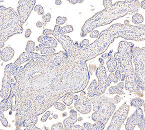 Immunohistochemical analysis of formalin fixed paraffin embedded human placenta tissue with F2400 at 1:50 dilution.