|
Come citare 1. Per la citazione nel testo (Materiali e Metodi): 2. Per la tabella delle risorse chiave: |
||
|
Numero verde: (877) 796-6397 -- Solo USA e Canada -- |
Fax: +1-832-582-8590 Ordini: +1-832-582-8158 |
Supporto tecnico: +1-832-582-8158 Ext:3 Si prega di fornire il numero dordine nelle-mail. Ci sforziamo di rispondere a tutte le richieste via e-mail entro un giorno lavorativo. |
Descrizione biologica
| Specificità | RBAP46/RBAP48 Antibody [P6E20] riconosce i livelli endogeni delle proteine totali RBAP46 e RBAP48. |
|---|---|
| Contesto | RbAp46 e RbAp48 (noti anche rispettivamente come RBBP7 e RBBP4) sono chaperoni istonici altamente simili che svolgono ruoli cruciali nella creazione e nel mantenimento della struttura della cromatina. Queste proteine sono componenti centrali di vari complessi coinvolti nell'aggiunta o nella rimozione di gruppi acetilici dai gruppi ɛ-amminici dei residui di lisina sulla coda N-terminale dell'istone H4 e sono anche parti vitali dei complessi di istone metiltransferasi. RbAp46 e RbAp48 si legano specificamente alla regione di ripiegamento dell'istone H4 e si ritiene che aiutino a dirigere i complessi di rimodellamento della cromatina, acetilazione e deacetilazione degli istoni ai loro substrati istonici. RbAp46 si lega specificamente e migliora l'attività dell'istone acetiltransferasi di tipo B HAT1, che acetila l'istone H4 ai residui di lisina 5 e 12 prima che questi istoni vengano incorporati nei nucleosomi durante la replicazione del DNA. RbAp48, d'altra parte, è una subunità conservata evolutivamente del complesso fattore-1 di assemblaggio della cromatina (CAF-1), dove interagisce con altre due subunità, p150 e p60, nelle cellule umane. Sia RbAp46 che RbAp48 sono coinvolti in numerosi altri complessi proteici, a volte coesistendo all'interno dello stesso complesso. Sono anche presenti nel complesso del fattore di rimodellamento dei nucleosomi (NURF), nel complesso di rimodellamento e deacetilazione dei nucleosomi (NuRD) e nel complesso di deacetilazione degli istoni Sin3/HDAC. |
Informazioni sullutilizzo
| Applicazione | WB | Diluizione |
|
||
|---|---|---|---|---|---|
| Reattività | Human, Mouse, Rat, Monkey | ||||
| Fonte | Rabbit Monoclonal Antibody | MW | 48 kDa | ||
| Tampone di conservazione | PBS, pH 7.2+50% Glycerol+0.05% BSA+0.01% NaN₃ | Conservazione (Dalla data di ricevimento) |
–20°C (avoid freeze-thaw cycles), 2 years | ||
| WB |
Experimental Protocol:
Sample preparation
1. Tissue: Lyse the tissue sample by adding an appropriate volume of ice-cold RIPA/Nuclear Lysis Buffer (containing Protease Inhibitor Cocktail),and homogenize the tissue at a low temperature or lyse it by sonication on ice, then incubate on ice for 30 minutes. 2. Adherent cell: Aspirate the culture medium and transfer the cells into an EP tube. Wash the cells with ice-cold PBS twice. Add an appropriate volume of RIPA/Nuclear Lysis Buffer (containing Protease Inhibitor Cocktail), sonicate to lyse the cells, and incubate on ice for 30 minutes. 3. Suspension cell: Transfer the culture medium to a pre-cooled centrifuge tube. Centrifuge and aspirate the supernatant. Wash the cells with ice-cold PBS twice.Add an appropriate volume of RIPA/Nuclear Lysis Buffer (containing Protease Inhibitor Cocktail), sonicate to lyse the cells, and incubate on ice for 30 minutes. 4. Place the lysate into a pre-cooled microcentrifuge tube. Centrifuge at 4°C for 15 min. Collect the supernatant;
5. Remove a small volume of lysate to determine the protein concentration;
6. Combine the lysate with protein loading buffer. Boil 20 µL sample under 95-100°C for 5 min. Centrifuge for 5 min after cool down on ice.
Electrophoretic separation
1. According to the concentration of extracted protein, load appropriate amount of protein sample and marker onto SDS-PAGE gels for electrophoresis. Recommended separating gel (lower gel) concentration: 10%. Reference Table for Selecting SDS-PAGE Separation Gel Concentrations 2. Power up 80V for 30 minutes. Then the power supply is adjusted (110 V~150 V), the Marker is observed, and the electrophoresis can be stopped when the indicator band of the predyed protein Marker where the protein is located is properly separated. (Note that the current should not be too large when electrophoresis, too large current (more than 150 mA) will cause the temperature to rise, affecting the result of running glue. If high currents cannot be avoided, an ice bath can be used to cool the bath.)
Transfer membrane
1. Take out the converter, soak the clip and consumables in the pre-cooled converter;
2. Activate PVDF membrane with methanol for 1 min and rinse with transfer buffer;
3. Install it in the order of "black edge of clip - sponge - filter paper - filter paper - glue -PVDF membrane - filter paper - filter paper - sponge - white edge of clip"; 4. The protein was electrotransferred to PVDF membrane. ( 0.45 µm PVDF membrane is recommended ) Reference Table for Selecting PVDF Membrane Pore Size Specifications Recommended conditions for wet transfer: 200 mA, 120 min. ( Note that the transfer conditions can be adjusted according to the protein size. For high-molecular-weight proteins, a higher current and longer transfer time are recommended. However, ensure that the transfer tank remains at a low temperature to prevent gel melting.)
Block
1. After electrotransfer, wash the film with TBST at room temperature for 5 minutes;
2. Incubate the film in the blocking solution for 1 hour at room temperature;
3. Wash the film with TBST for 3 times, 5 minutes each time.
Antibody incubation
1. Use 5% skim milk powder to prepare the primary antibody working liquid (recommended dilution ratio for primary antibody 1:1000), gently shake and incubate with the film at 4°C overnight; 2. Wash the film with TBST 3 times, 5 minutes each time;
3. Add the secondary antibody to the blocking solution and incubate with the film gently at room temperature for 1 hour;
4. After incubation, wash the film with TBST 3 times for 5 minutes each time.
Antibody staining
922. Add the prepared ECL luminescent substrate (or select other color developing substrate according to the second antibody) and mix evenly;
2. Incubate with the film for 1 minute, remove excess substrate (keep the film moist), wrap with plastic film, and expose in the imaging system.
|
Riferimenti
|
Dati di applicazione
WB
Validato da Selleck
-
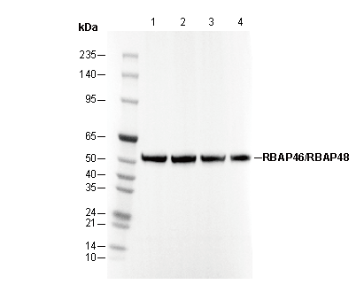 Lane 1: SH-SY5Y
Lane 2: COS-7
Lane 3: F9
Lane 4: H-4-II-E