|
Come citare 1. Per la citazione nel testo (Materiali e Metodi): 2. Per la tabella delle risorse chiave: |
||
|
Numero verde: (877) 796-6397 -- Solo USA e Canada -- |
Fax: +1-832-582-8590 Ordini: +1-832-582-8158 |
Supporto tecnico: +1-832-582-8158 Ext:3 Si prega di fornire il numero dordine nelle-mail. Ci sforziamo di rispondere a tutte le richieste via e-mail entro un giorno lavorativo. |
Descrizione biologica
| Specificità | PI3 Kinase p110 δ Antibody [H17B15] riconosce i livelli endogeni della proteina PI3 Kinase Class III δ totale. |
|---|---|
| Contesto | La fosfoinositide 3-chinasi (PI3K) è un enzima che fosforila il fosfatidilinositolo (PI), il fosfatidilinositolo-4-fosfato (PIP) e il fosfatidilinositolo-4,5-bisfosfato (PIP2) per produrre fosfatidilinositolo-3,4,5-trifosfato. Questo processo è attivato da fattori di crescita e ormoni, portando alla regolazione della crescita cellulare, della progressione del ciclo cellulare, della migrazione cellulare e della sopravvivenza. L'enzima PTEN contrasta questo processo defosforilando il fosfatidilinositolo-3,4,5-trifosfato, e gli studi hanno dimostrato che la perdita della funzione di PTEN si traduce in un'attivazione costitutiva della via PI3K in vari tumori umani. Le PI3K sono costituite da una subunità catalitica (p110) e una subunità regolatrice, con diverse isoforme della subunità catalitica, tra cui p110α, p110β, p110γ e p110δ. Le subunità regolatrice p85α e p85β interagiscono con p110α, p110β e p110δ. Sebbene le mutazioni con guadagno di funzione nel gene PIK3CA, che codifica l'isoforma p110α, siano comunemente osservate in molti tumori umani, non sono state trovate mutazioni somatiche nei geni che codificano per p110β o p110δ. A differenza delle ampiamente espresse p110α e p110β, la p110δ si trova prevalentemente nei leucociti, rendendo la via p110δ un focus di ricerca nei disturbi immunitari. L'isoforma p110δ ha un ruolo chiave nello sviluppo e nella progressione di alcune neoplasie ematologiche, con studi che utilizzano inibitori selettivi di p110δ e inattivazione genetica in modelli murini che evidenziano il suo coinvolgimento in processi come la differenziazione cellulare, la crescita, la sopravvivenza, la motilità e l'interazione con l'inositolo fosfatasi PTEN. |
Informazioni sullutilizzo
| Applicazione | WB, IP | Diluizione |
|
||||
|---|---|---|---|---|---|---|---|
| Reattività | Human, Mouse, Rat | ||||||
| Fonte | Rabbit Monoclonal Antibody | MW | 110 kDa | ||||
| Tampone di conservazione | PBS, pH 7.2+50% Glycerol+0.05% BSA+0.01% NaN₃ | Conservazione (Dalla data di ricevimento) |
-20°C (avoid freeze-thaw cycles), 2 years | ||||
| WB |
Experimental Protocol:
Sample preparation
1. Tissue: Lyse the tissue sample by adding an appropriate volume of ice-cold RIPA/Tris-HCL Lysis Buffer (containing Protease Inhibitor Cocktail),and homogenize the tissue at a low temperature or lyse it by sonication on ice, then incubate on ice for 30 minutes. 2. Adherent cell: Aspirate the culture medium and transfer the cells into an EP tube. Wash the cells with ice-cold PBS twice. Add an appropriate volume of RIPA/Tris-HCL Lysis Buffer (containing Protease Inhibitor Cocktail), sonicate to lyse the cells, and incubate on ice for 30 minutes. 3. Suspension cell: Transfer the culture medium to a pre-cooled centrifuge tube. Centrifuge and aspirate the supernatant. Wash the cells with ice-cold PBS twice.Add an appropriate volume of RIPA/Tris-HCL Lysis Buffer (containing Protease Inhibitor Cocktail), sonicate to lyse the cells, and incubate on ice for 30 minutes. 4. Place the lysate into a pre-cooled microcentrifuge tube. Centrifuge at 4°C for 15 min. Collect the supernatant;
5. Remove a small volume of lysate to determine the protein concentration;
6. Combine the lysate with protein loading buffer. Boil 20 µL sample under 95-100°C for 5 min. Centrifuge for 5 min after cool down on ice.
Electrophoretic separation
1. According to the concentration of extracted protein, load appropriate amount of protein sample and marker onto SDS-PAGE gels for electrophoresis. Recommended separating gel (lower gel) concentration: 5%. Reference Table for Selecting SDS-PAGE Separation Gel Concentrations 2. Power up 80V for 30 minutes. Then the power supply is adjusted (110 V~150 V), the Marker is observed, and the electrophoresis can be stopped when the indicator band of the predyed protein Marker where the protein is located is properly separated. (Note that the current should not be too large when electrophoresis, too large current (more than 150 mA) will cause the temperature to rise, affecting the result of running glue. If high currents cannot be avoided, an ice bath can be used to cool the bath.)
Transfer membrane
1. Take out the converter, soak the clip and consumables in the pre-cooled converter;
2. Activate PVDF membrane with methanol for 1 min and rinse with transfer buffer;
3. Install it in the order of "black edge of clip - sponge - filter paper - filter paper - glue -PVDF membrane - filter paper - filter paper - sponge - white edge of clip"; 4. The protein was electrotransferred to PVDF membrane. ( 0.45 µm PVDF membrane is recommended ) Reference Table for Selecting PVDF Membrane Pore Size Specifications Recommended conditions for wet transfer: 200 mA, 120 min. ( Note that the transfer conditions can be adjusted according to the protein size. For high-molecular-weight proteins, a higher current and longer transfer time are recommended. However, ensure that the transfer tank remains at a low temperature to prevent gel melting.)
Block
1. After electrotransfer, wash the film with TBST at room temperature for 5 minutes;
2. Incubate the film in the blocking solution for 1 hour at room temperature;
3. Wash the film with TBST for 3 times, 5 minutes each time.
Antibody incubation
1. Use 5% skim milk powder to prepare the primary antibody working liquid (recommended dilution ratio for primary antibody 1:1000), gently shake and incubate with the film at 4°C overnight; 2. Wash the film with TBST 3 times, 5 minutes each time;
3. Add the secondary antibody to the blocking solution and incubate with the film gently at room temperature for 1 hour;
4. After incubation, wash the film with TBST 3 times for 5 minutes each time.
Antibody staining
1128. Add the prepared ECL luminescent substrate (or select other color developing substrate according to the second antibody) and mix evenly;
2. Incubate with the film for 1 minute, remove excess substrate (keep the film moist), wrap with plastic film, and expose in the imaging system.
|
Riferimenti
|
Dati di applicazione
WB
Validato da Selleck
-
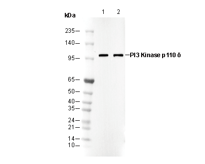 Lane 1: SW620
Lane 2: HT1080