|
Come citare 1. Per la citazione nel testo (Materiali e Metodi): 2. Per la tabella delle risorse chiave: |
||
|
Numero verde: (877) 796-6397 -- Solo USA e Canada -- |
Fax: +1-832-582-8590 Ordini: +1-832-582-8158 |
Supporto tecnico: +1-832-582-8158 Ext:3 Si prega di fornire il numero dordine nelle-mail. Ci sforziamo di rispondere a tutte le richieste via e-mail entro un giorno lavorativo. |
Descrizione biologica
| Specificità | Phospho-Tau (Ser202/Thr205) Antibody [M2L8] riconosce i livelli endogeni della proteina Tau totale solo quando fosforilata sia in Ser202 che in Thr205. |
|---|---|
| Contesto | Phospho-Tau (Ser202/Thr205) si riferisce alla proteina tau fosforilata in serina 202 e treonina 205, due residui adiacenti all'interno della regione ricca di prolina di questa proteina Microtubule Associated. Tau è una proteina intrinsecamente disordinata che stabilizza e promuove l'assemblaggio dei microtubuli. La fosforilazione in Ser202/Thr205 induce un cambiamento conformazionale che estende la regione N-terminale e aumenta la spaziatura tra i microtubuli nei neuriti. Questi siti sono fosforilati da chinasi come Cdk5, GSK-3β, PKA e caseina chinasi II, con Cdk5 che fosforila preferenzialmente Thr205 prima di Ser202, e l'ordine di fosforilazione influenzato dall'associazione di tau con i microtubuli. La fosforilazione in questi siti regola l'affinità di legame di tau ai microtubuli, riducendo generalmente la sua capacità di stabilizzare i microtubuli e contribuendo alla cytoskeletal destabilizzazione. L'iperfosforilazione in Ser202/Thr205 è un evento chiave nelle malattie neurodegenerative, che porta all'aggregazione di tau e alla formazione di grovigli neurofibrillari, che sono caratteristici della malattia di Alzheimer e di altre tauopatie. Lo stato di fosforilazione di questi residui è anche modulato da varie fosfatasi, tra cui PP2A, PP2B e PP5, che sono più efficaci di PP1 nel defosforilare tau in questi siti. Queste modificazioni alterano la conformazione di tau, le interazioni con i microtubuli e la localizzazione cellulare, e svolgono un ruolo centrale nella progressione della malattia compromettendo la funzione neuronale e promuovendo la patologia di tau. |
Informazioni sullutilizzo
| Applicazione | WB | Diluizione |
|
||
|---|---|---|---|---|---|
| Reattività | Human, Mouse, Rat | ||||
| Fonte | Rabbit Monoclonal Antibody | MW | 55-80 kDa | ||
| Tampone di conservazione | PBS, pH 7.2+50% Glycerol+0.05% BSA+0.01% NaN3 | Conservazione (Dalla data di ricevimento) |
-20°C (avoid freeze-thaw cycles), 2 years | ||
| WB |
Experimental Protocol:
Sample preparation
1. Tissue: Lyse the tissue sample by adding an appropriate volume of ice-cold RIPA/NP-40 Lysis Buffer (containing Protease Inhibitor Cocktail, Phosphatase Inhibitor Cocktail),and homogenize the tissue at a low temperature. 2. Adherent cell: Aspirate the culture medium and wash the cells with ice-cold PBS twice. Lyse the cells by adding an appropriate volume of RIPA/NP-40 Lysis Buffer (containing Protease Inhibitor Cocktail, Phosphatase Inhibitor Cocktail) and put the sample on ice for 5 min. 3. Suspension cell: Transfer the culture medium to a pre-cooled centrifuge tube. Centrifuge and aspirate the supernatant. Wash the cells with ice-cold PBS twice. Lyse the cells by adding an appropriate volume of RIPA/NP-40 Lysis Buffer (containing Protease Inhibitor Cocktail, Phosphatase Inhibitor Cocktail) and put the sample on ice for 5 min. 4. Place the lysate into a pre-cooled microcentrifuge tube. Centrifuge at 4°C for 15 min. Collect the supernatant;
5. Remove a small volume of lysate to determine the protein concentration;
6. Combine the lysate with protein loading buffer. Boil 20 µL sample under 95-100°C for 5 min. Centrifuge for 5 min after cool down on ice.
Electrophoretic separation
1. According to the concentration of extracted protein, load appropriate amount of protein sample and marker onto SDS-PAGE gels for electrophoresis. Recommended separating gel (lower gel) concentration: 10%. Reference Table for Selecting SDS-PAGE Separation Gel Concentrations 2. Power up 80V for 30 minutes. Then the power supply is adjusted (110 V~150 V), the Marker is observed, and the electrophoresis can be stopped when the indicator band of the predyed protein Marker where the protein is located is properly separated. (Note that the current should not be too large when electrophoresis, too large current (more than 150 mA) will cause the temperature to rise, affecting the result of running glue. If high currents cannot be avoided, an ice bath can be used to cool the bath.)
Transfer membrane
1. Take out the converter, soak the clip and consumables in the pre-cooled converter;
2. Activate PVDF membrane with methanol for 1 min and rinse with transfer buffer;
3. Install it in the order of "black edge of clip - sponge - filter paper - filter paper - glue -PVDF membrane - filter paper - filter paper - sponge - white edge of clip"; 4. The protein was electrotransferred to PVDF membrane. ( 0.45 µm PVDF membrane is recommended ) Reference Table for Selecting PVDF Membrane Pore Size Specifications Recommended conditions for wet transfer: 200 mA, 120 min. ( Note that the transfer conditions can be adjusted according to the protein size. For high-molecular-weight proteins, a higher current and longer transfer time are recommended. However, ensure that the transfer tank remains at a low temperature to prevent gel melting.)
Block
1. After electrotransfer, wash the film with TBST at room temperature for 5 minutes;
2. Incubate the film in the blocking solution ( recommending 5% BSA solution)
for 1 hour at room temperature;
3. Wash the film with TBST for 3 times, 5 minutes each time.
Antibody incubation
1. Use 5% skim milk powder to prepare the primary antibody working liquid (recommended dilution ratio for primary antibody 1:1000), gently shake and incubate with the film at 4°C overnight; 2. Wash the film with TBST 3 times, 5 minutes each time;
3. Add the secondary antibody to the blocking solution and incubate with the film gently at room temperature for 1 hour;
4. After incubation, wash the film with TBST 3 times for 5 minutes each time.
Antibody staining
1. Add the prepared ECL luminescent substrate (or select other color developing substrate according to the second antibody) and mix evenly;
2. Incubate with the film for 1 minute, remove excess substrate (keep the film moist), wrap with plastic film, and expose in the imaging system. (Exposure time of at least 120s is recommended)
|
Riferimenti
|
Dati di applicazione
WB
Validato da Selleck
-
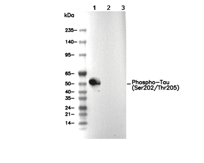 Lane 1: Mouse brain, Lane 2: Mouse brain (λ phosphatase-treated), Lane 3: Mouse brain (KO Tau)