|
Come citare 1. Per la citazione nel testo (Materiali e Metodi): 2. Per la tabella delle risorse chiave: |
||
|
Numero verde: (877) 796-6397 -- Solo USA e Canada -- |
Fax: +1-832-582-8590 Ordini: +1-832-582-8158 |
Supporto tecnico: +1-832-582-8158 Ext:3 Si prega di fornire il numero dordine nelle-mail. Ci sforziamo di rispondere a tutte le richieste via e-mail entro un giorno lavorativo. |
Descrizione biologica
| Specificità | Phospho-RIP3 (Ser232) Antibody [E19K17] riconosce i livelli endogeni della proteina RIP3 totale solo quando fosforilata a Ser232 |
|---|---|
| Contesto | La famiglia delle serina/treonina chinasi interagenti con i recettori (RIP) – inclusi RIP1, RIP2, RIP3 e RIP4 – svolge un ruolo cruciale nella modulazione delle risposte cellulari allo stress. Queste chinasi sono coinvolte nell'avvio di segnali pro-sopravvivenza e infiammatori, principalmente attraverso l'attivazione di NF-κB, nonché nella promozione di vie di morte cellulare come Apoptosis. Tra queste, la receptor-interacting protein kinase 3 (RIPK3 o RIP3) è un attore centrale nella necroptosi, una forma regolata di morte cellulare necrotica. RIP3 partecipa a cascate di segnalazione avviate dal fattore di necrosi tumorale (TNF), dove si associa a RIP1 e al complesso del recettore TNF per mediare sia l'attivazione di NF-κB che Apoptosis. Un evento chiave nella necroptosi è l'interazione tra RIP1 e RIP3, che costituisce la base del necrosoma – un complesso multiproteico che facilita la necrosi programmata. Questa morte cellulare di tipo necrotico è tipicamente innescata dalla segnalazione di TNF in presenza di inibizione della caspasi. Negli esseri umani, la fosforilazione di RIP3 alla Serina 227 è critica per il suo legame con la proteina simile al dominio chinasi a linea mista (MLKL), consentendo l'assemblaggio del necrosoma. Nei topi, la stimolazione del TNF induce la fosforilazione alla Treonina 231 e Serina 232 su RIP3, necessaria per la sua interazione con la MLKL murina. In particolare, la Ser-232 nella RIP3 murina corrisponde alla Ser-227 nella proteina umana. Inoltre, RIP1 e RIP3 contribuiscono all'oligomerizzazione del necrosoma, che promuove la formazione di strutture di segnalazione simili all'amiloide essenziali per l'esecuzione della necroptosi. |
Informazioni sullutilizzo
| Applicazione | WB, ELISA | Diluizione |
|
||
|---|---|---|---|---|---|
| Reattività | Mouse | ||||
| Fonte | Rabbit Monoclonal Antibody | MW | 53 kDa | ||
| Tampone di conservazione | PBS, pH 7.2+50% Glycerol+0.05% BSA+0.01% NaN3 | Conservazione (Dalla data di ricevimento) |
-20°C (avoid freeze-thaw cycles), 2 years | ||
| WB |
Experimental Protocol:
Sample preparation
1. Tissue: Lyse the tissue sample by adding an appropriate volume of ice-cold RIPA/Nuclear Lysis Buffer (containing Protease Inhibitor Cocktail, Phosphatase Inhibitor Cocktail),and homogenize the tissue at a low temperature. 2. Adherent cell: Aspirate the culture medium and wash the cells with ice-cold PBS twice. Lyse the cells by adding an appropriate volume of RIPA/Nuclear Lysis Buffer (containing Protease Inhibitor Cocktail, Phosphatase Inhibitor Cocktail) and put the sample on ice for 5 min. 3. Suspension cell: Transfer the culture medium to a pre-cooled centrifuge tube. Centrifuge and aspirate the supernatant. Wash the cells with ice-cold PBS twice. Lyse the cells by adding an appropriate volume of RIPA/Nuclear Lysis Buffer (containing Protease Inhibitor Cocktail, Phosphatase Inhibitor Cocktail) and put the sample on ice for 5 min. 4. Place the lysate into a pre-cooled microcentrifuge tube. Centrifuge at 4°C for 15 min. Collect the supernatant;
5. Remove a small volume of lysate to determine the protein concentration;
6. Combine the lysate with protein loading buffer. Boil 20 µL sample under 95-100°C for 5 min. Centrifuge for 5 min after cool down on ice.
Electrophoretic separation
1. According to the concentration of extracted protein, load appropriate amount of protein sample and marker onto SDS-PAGE gels for electrophoresis. Recommended separating gel (lower gel) concentration: 10%. Reference Table for Selecting SDS-PAGE Separation Gel Concentrations 2. Power up 80V for 30 minutes. Then the power supply is adjusted (110 V~150 V), the Marker is observed, and the electrophoresis can be stopped when the indicator band of the predyed protein Marker where the protein is located is properly separated. (Note that the current should not be too large when electrophoresis, too large current (more than 150 mA) will cause the temperature to rise, affecting the result of running glue. If high currents cannot be avoided, an ice bath can be used to cool the bath.)
Transfer membrane
1. Take out the converter, soak the clip and consumables in the pre-cooled converter;
2. Activate PVDF membrane with methanol for 1 min and rinse with transfer buffer;
3. Install it in the order of "black edge of clip - sponge - filter paper - filter paper - glue -PVDF membrane - filter paper - filter paper - sponge - white edge of clip"; 4. The protein was electrotransferred to PVDF membrane. ( 0.45 µm PVDF membrane is recommended ) Reference Table for Selecting PVDF Membrane Pore Size Specifications Recommended conditions for wet transfer: 200 mA, 120 min. ( Note that the transfer conditions can be adjusted according to the protein size. For high-molecular-weight proteins, a higher current and longer transfer time are recommended. However, ensure that the transfer tank remains at a low temperature to prevent gel melting.)
Block
1. After electrotransfer, wash the film with TBST at room temperature for 5 minutes;
2. Incubate the film in the blocking solution ( recommending 5% BSA solution)
for 1 hour at room temperature;
3. Wash the film with TBST for 3 times, 5 minutes each time.
Antibody incubation
1. Use 5% skim milk powder to prepare the primary antibody working liquid (recommended dilution ratio for primary antibody 1:1000), gently shake and incubate with the film at 4°C overnight; 2. Wash the film with TBST 3 times, 5 minutes each time;
3. Add the secondary antibody to the blocking solution and incubate with the film gently at room temperature for 1 hour;
4. After incubation, wash the film with TBST 3 times for 5 minutes each time.
Antibody staining
1. Add the prepared ECL luminescent substrate (or select other color developing substrate according to the second antibody) and mix evenly;
2. Incubate with the film for 1 minute, remove excess substrate (keep the film moist), wrap with plastic film, and expose in the imaging system.
|
Riferimenti
|
Dati di applicazione
WB
Validato da Selleck
-
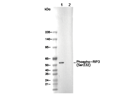 Lane 1: L-929, Lane 2: L-929 (phosphatase-treated)