|
Come citare 1. Per la citazione nel testo (Materiali e Metodi): 2. Per la tabella delle risorse chiave: |
||
|
Numero verde: (877) 796-6397 -- Solo USA e Canada -- |
Fax: +1-832-582-8590 Ordini: +1-832-582-8158 |
Supporto tecnico: +1-832-582-8158 Ext:3 Si prega di fornire il numero dordine nelle-mail. Ci sforziamo di rispondere a tutte le richieste via e-mail entro un giorno lavorativo. |
Descrizione biologica
| Specificità | Phospho-Met (Tyr1349) Antibody [D24L22] rileva i livelli endogeni della proteina Met totale solo quando è fosforilata in Tyr1349. |
|---|---|
| Contesto | Phospho-Met (Tyr1349) si riferisce allo stato attivato del recettore del fattore di crescita degli epatociti Met, una tirosin-chinasi recettoriale eterodimerica legata da disolfuro (catena α extracellulare di ~45 kDa e catena β transmembrana di ~145 kDa) che si lega all'HGF per orchestrare programmi di crescita invasivi come la proliferazione, la sopravvivenza, la migrazione e la morfogenesi nelle cellule epiteliali e in altri tipi cellulari. Met presenta un dominio semaforina (SEMA), PSI e quattro ripetizioni IPT nella sua regione extracellulare per il riconoscimento dell'HGF e la dimerizzazione, una singola elica transmembrana e un segmento intracellulare con una regione iuxtamembrana (contenente Tyr1003 per l'ubiquitinazione/degradazione mediata da CBL e Ser957/Ser975 inibitorie), un dominio tirosin-chinasi (con Tyr1234/Tyr1235 dell'ansa di attivazione la cui fosforilazione migliora l'attività catalitica) e una coda C-terminale che ospita Tyr1349 e Tyr1356 che formano un sito di attracco multisubstrato. Dopo il legame con HGF, Met dimerizza e il dominio chinasico transfosforila Tyr1234/1235 per stabilizzare la conformazione attiva, consentendo la successiva fosforilazione di Tyr1349 (e Tyr1356). Phospho-Tyr1349 fornisce un sito di legame ad alta affinità per l'adattatore Gab1 e recluta effettori contenenti SH2, inclusi GRB2, PI3K p85, SHP2, Src e STAT3, assemblando un hub di segnalazione che connette Met alle vie Ras-MAPK, PI3K-Akt, STAT e GTPasi della famiglia Rho per guidare la progressione del ciclo cellulare, la sopravvivenza, la motilità e la transizione epitelio-mesenchimale. La mutazione o la perdita di Tyr1349/Tyr1356 compromette l'attracco di Gab1 e l'attivazione a valle di PI3K/MAPK nonostante l'attività chinasica preservata. |
Informazioni sullutilizzo
| Applicazione | WB | Diluizione |
|
||
|---|---|---|---|---|---|
| Reattività | Human, Mouse, Rat | ||||
| Fonte | Rabbit Monoclonal Antibody | MW | 145 kDa | ||
| Tampone di conservazione | PBS, pH 7.2+50% Glycerol+0.05% BSA+0.01% NaN3 | Conservazione (Dalla data di ricevimento) |
-20°C (avoid freeze-thaw cycles), 2 years | ||
| WB |
Experimental Protocol:
Sample preparation
1. Tissue: Lyse the tissue sample by adding an appropriate volume of ice-cold RIPA/NP-40 Lysis Buffer (containing Protease Inhibitor Cocktail, Phosphatase Inhibitor Cocktail),and homogenize the tissue at a low temperature. 2. Adherent cell: Aspirate the culture medium and wash the cells with ice-cold PBS twice. Lyse the cells by adding an appropriate volume of RIPA/NP-40 Lysis Buffer (containing Protease Inhibitor Cocktail, Phosphatase Inhibitor Cocktail) and put the sample on ice for 5 min. 3. Suspension cell: Transfer the culture medium to a pre-cooled centrifuge tube. Centrifuge and aspirate the supernatant. Wash the cells with ice-cold PBS twice. Lyse the cells by adding an appropriate volume of RIPA/NP-40 Lysis Buffer (containing Protease Inhibitor Cocktail, Phosphatase Inhibitor Cocktail) and put the sample on ice for 5 min. 4. Place the lysate into a pre-cooled microcentrifuge tube. Centrifuge at 4°C for 15 min. Collect the supernatant;
5. Remove a small volume of lysate to determine the protein concentration;
6. Combine the lysate with protein loading buffer. Boil 20 µL sample under 95-100°C for 5 min. Centrifuge for 5 min after cool down on ice.
Electrophoretic separation
1. According to the concentration of extracted protein, load appropriate amount of protein sample and marker onto SDS-PAGE gels for electrophoresis. Recommended separating gel (lower gel) concentration: 5%. Reference Table for Selecting SDS-PAGE Separation Gel Concentrations 2. Power up 80V for 30 minutes. Then the power supply is adjusted (110 V~150 V), the Marker is observed, and the electrophoresis can be stopped when the indicator band of the predyed protein Marker where the protein is located is properly separated. (Note that the current should not be too large when electrophoresis, too large current (more than 150 mA) will cause the temperature to rise, affecting the result of running glue. If high currents cannot be avoided, an ice bath can be used to cool the bath.)
Transfer membrane
1. Take out the converter, soak the clip and consumables in the pre-cooled converter;
2. Activate PVDF membrane with methanol for 1 min and rinse with transfer buffer;
3. Install it in the order of "black edge of clip - sponge - filter paper - filter paper - glue -PVDF membrane - filter paper - filter paper - sponge - white edge of clip"; 4. The protein was electrotransferred to PVDF membrane. ( 0.45 µm PVDF membrane is recommended ) Reference Table for Selecting PVDF Membrane Pore Size Specifications Recommended conditions for wet transfer: 200 mA, 120 min. ( Note that the transfer conditions can be adjusted according to the protein size. For high-molecular-weight proteins, a higher current and longer transfer time are recommended. However, ensure that the transfer tank remains at a low temperature to prevent gel melting.)
Block
1. After electrotransfer, wash the film with TBST at room temperature for 5 minutes;
2. Incubate the film in the blocking solution ( recommending 5% BSA solution)
for 1 hour at room temperature;
3. Wash the film with TBST for 3 times, 5 minutes each time.
Antibody incubation
1. Use 5% skim milk powder to prepare the primary antibody working liquid (recommended dilution ratio for primary antibody 1:1000), gently shake and incubate with the film at 4°C overnight; 2. Wash the film with TBST 3 times, 5 minutes each time;
3. Add the secondary antibody to the blocking solution and incubate with the film gently at room temperature for 1 hour;
4. After incubation, wash the film with TBST 3 times for 5 minutes each time.
Antibody staining
1. Add the prepared ECL luminescent substrate (or select other color developing substrate according to the second antibody) and mix evenly;
2. Incubate with the film for 1 minute, remove excess substrate (keep the film moist), wrap with plastic film, and expose in the imaging system.
|
Riferimenti
|
Dati di applicazione
WB
Validato da Selleck
-
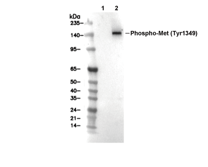 Lane 1: H-4-II-E, Lane 2: H-4-II-E (HGF, 50ng/mL, 5min)