|
Come citare 1. Per la citazione nel testo (Materiali e Metodi): 2. Per la tabella delle risorse chiave: |
||
|
Numero verde: (877) 796-6397 -- Solo USA e Canada -- |
Fax: +1-832-582-8590 Ordini: +1-832-582-8158 |
Supporto tecnico: +1-832-582-8158 Ext:3 Si prega di fornire il numero dordine nelle-mail. Ci sforziamo di rispondere a tutte le richieste via e-mail entro un giorno lavorativo. |
Descrizione biologica
| Specificità | Phospho-IκBα (Ser32/36) Antibody [F24N7] rileva i livelli endogeni di IκBα solo quando fosforilato a Ser32/36. |
|---|---|
| Contesto | Il complesso IκB chinasi (IKK) funge da hub di segnalazione centrale per l'attivazione della via NF-κB. Questo complesso è composto da due subunità catalitiche, IKKα e IKKβ, che sono serina/treonina chinasi, e una subunità regolatrice nota come NEMO (NF-κB essential modulator), anche definita IKKγ. Il complesso IKK integra una vasta gamma di segnali a monte che attivano NF-κB, portando alla fosforilazione delle proteine IκB, delle subunità NF-κB e di altri bersagli cellulari. Nelle cellule non stimolate, i fattori di trascrizione NF-κB sono mantenuti in uno stato inattivo nel citoplasma legandosi a proteine IκB inibitorie, come IκBα, IκBβ e IκBε, o alle proteine precursori p100 e p105. Queste proteine inibitorie impediscono a NF-κB di entrare nel nucleo e di iniziare la trascrizione genica. In seguito a stimolazione, le proteine IκB vengono fosforilate a residui di serina specifici — in particolare Ser32 e Ser36 nel caso di IκBα — dal complesso IKK attivato. Questa fosforilazione contrassegna IκB per il riconoscimento da parte del complesso di ubiquitina ligasi E3 SCFβTrCP, portando alla sua ubiquitinazione e successiva degradazione tramite il proteasoma. La degradazione di IκB rilascia i dimeri NF-κB, che poi traslocano nel nucleo dove guidano la trascrizione dei geni bersaglio. Una vasta varietà di segnali esterni, inclusi citochine infiammatorie, fattori di crescita e chemochine, può innescare questa cascata, risultando in ultima analisi nella fosforilazione di IκBα e nell'attivazione dell'espressione genica dipendente da NF-κB. |
Informazioni sullutilizzo
| Applicazione | WB | Diluizione |
|
||
|---|---|---|---|---|---|
| Reattività | Human, Mouse, Rat, Monkey | ||||
| Fonte | Mouse Monoclonal Antibody | MW | 40 kDa | ||
| Tampone di conservazione | PBS, pH 7.2+50% Glycerol+0.05% BSA+0.01% NaN3 | Conservazione (Dalla data di ricevimento) |
-20°C (avoid freeze-thaw cycles), 2 years | ||
| WB |
Experimental Protocol:
Sample preparation
1. Tissue: Lyse the tissue sample by adding an appropriate volume of ice-cold RIPA/Nuclear Lysis Buffer (containing Protease Inhibitor Cocktail, Phosphatase Inhibitor Cocktail),and homogenize the tissue at a low temperature. 2. Adherent cell: Aspirate the culture medium and wash the cells with ice-cold PBS twice. Lyse the cells by adding an appropriate volume of RIPA/Nuclear Lysis Buffer (containing Protease Inhibitor Cocktail, Phosphatase Inhibitor Cocktail) and put the sample on ice for 5 min. 3. Suspension cell: Transfer the culture medium to a pre-cooled centrifuge tube. Centrifuge and aspirate the supernatant. Wash the cells with ice-cold PBS twice. Lyse the cells by adding an appropriate volume of RIPA/Nuclear Lysis Buffer (containing Protease Inhibitor Cocktail, Phosphatase Inhibitor Cocktail) and put the sample on ice for 5 min. 4. Place the lysate into a pre-cooled microcentrifuge tube. Centrifuge at 4°C for 15 min. Collect the supernatant;
5. Remove a small volume of lysate to determine the protein concentration;
6. Combine the lysate with protein loading buffer. Boil 20 µL sample under 95-100°C for 5 min. Centrifuge for 5 min after cool down on ice.
Electrophoretic separation
1. According to the concentration of extracted protein, load appropriate amount of protein sample and marker onto SDS-PAGE gels for electrophoresis. Recommended separating gel (lower gel) concentration: 10%. Reference Table for Selecting SDS-PAGE Separation Gel Concentrations 2. Power up 80V for 30 minutes. Then the power supply is adjusted (110 V~150 V), the Marker is observed, and the electrophoresis can be stopped when the indicator band of the predyed protein Marker where the protein is located is properly separated. (Note that the current should not be too large when electrophoresis, too large current (more than 150 mA) will cause the temperature to rise, affecting the result of running glue. If high currents cannot be avoided, an ice bath can be used to cool the bath.)
Transfer membrane
1. Take out the converter, soak the clip and consumables in the pre-cooled converter;
2. Activate PVDF membrane with methanol for 1 min and rinse with transfer buffer;
3. Install it in the order of "black edge of clip - sponge - filter paper - filter paper - glue -PVDF membrane - filter paper - filter paper - sponge - white edge of clip"; 4. The protein was electrotransferred to PVDF membrane. ( 0.45 µm PVDF membrane is recommended ) Reference Table for Selecting PVDF Membrane Pore Size Specifications Recommended conditions for wet transfer: 200 mA, 60 min. ( Note that the transfer conditions can be adjusted according to the protein size. For high-molecular-weight proteins, a higher current and longer transfer time are recommended. However, ensure that the transfer tank remains at a low temperature to prevent gel melting.)
Block
1. After electrotransfer, wash the film with TBST at room temperature for 5 minutes;
2. Incubate the film in the blocking solution ( recommending 5% BSA solution)
for 1 hour at room temperature;
3. Wash the film with TBST for 3 times, 5 minutes each time.
Antibody incubation
1. Use 5% skim milk powder to prepare the primary antibody working liquid (recommended dilution ratio for primary antibody 1:1000), gently shake and incubate with the film at 4°C overnight; 2. Wash the film with TBST 3 times, 5 minutes each time;
3. Add the secondary antibody to the blocking solution and incubate with the film gently at room temperature for 1 hour;
4. After incubation, wash the film with TBST 3 times for 5 minutes each time.
Antibody staining
1. Add the prepared ECL luminescent substrate (or select other color developing substrate according to the second antibody) and mix evenly;
2. Incubate with the film for 1 minute, remove excess substrate (keep the film moist), wrap with plastic film, and expose in the imaging system. (Exposure time of at least 150s is recommended)
|
Riferimenti
|
Dati di applicazione
WB
Validato da Selleck
-
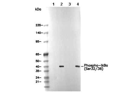 Lane 1: Hela, Lane 2: Hela (IFNa, 20 ng/mL, 5 min), Lane 3: 3T3, Lane 4: 3T3 (IFNa, 20 ng/mL, 5 min)