|
Come citare 1. Per la citazione nel testo (Materiali e Metodi): 2. Per la tabella delle risorse chiave: |
||
|
Numero verde: (877) 796-6397 -- Solo USA e Canada -- |
Fax: +1-832-582-8590 Ordini: +1-832-582-8158 |
Supporto tecnico: +1-832-582-8158 Ext:3 Si prega di fornire il numero dordine nelle-mail. Ci sforziamo di rispondere a tutte le richieste via e-mail entro un giorno lavorativo. |
Descrizione biologica
| Specificità | Phospho-Chk1 (Ser296) Antibody [F19D13] rileva i livelli endogeni della proteina Chk1 totale solo quando è fosforilata in Ser296. |
|---|---|
| Contesto | Phospho-Chk1 (Ser296) rappresenta la forma attivata della chinasi checkpoint 1 (Chk1), una serina/treonina chinasi cruciale per la risposta al danno del DNA (DDR) che integra la segnalazione ATR/ATM per rafforzare i checkpoint del ciclo cellulare, stabilizzare le forcelle di replicazione e promuovere la riparazione del DNA o l'apoptosi. Chk1 è una proteina con un dominio chinasico N-terminale contenente la triade catalitica conservata (Lys38, Glu91, Asp147 nell'ansa di attivazione) e una coda C-terminale regolatoria con siti SQ (Ser317, Ser345) per la fosforilazione da parte di ATR, oltre a un sito di autofosforilazione in Ser296 situato in un'ansa non strutturata. La fosforilazione in Ser296 avviene tramite un meccanismo di autofosforilazione cis-intramolecolare dipendente da ATR solo dopo la fosforilazione di priming in Ser317/Ser345, eventi che alleviano l'autoinibizione, stabilizzano la conformazione chinasica attiva ed espongono Ser296 per l'autofosforilazione. Phospho-Ser296 Chk1 migliora l'efficienza del checkpoint aumentando l'attività chinasica verso le fosfatasi Cdc25 (Cdc25A Ser76 per la proteolisi mediata da β-TrCP, Cdc25B/C Ser216/287 per la sequestrazione 14-3-3), prevenendo così l'attivazione prematura di CDK1/2 e promuovendo l'arresto del Cell Cycle in S/G2/M. Fosforila anche effettori aggiuntivi come Treslin, Claspin e FanD2 per coordinare il riavvio della forcella di replicazione, la ricombinazione omologa e la sintesi di traslesione, mentre la phospho-Chk1 centrosomale inibisce Cdc25B-CDK1 per bloccare l'ingresso mitotico fino a quando il danno al DNA non è risolto. Questa cascata di attivazione multi-sito assicura una robusta segnalazione DDR, e i mutanti Ser296A mostrano un arresto del checkpoint difettoso e letalità sintetica con gli inibitori di ATR. Phospho-Chk1(Ser296) indica lo stress replicativo e predice la sensibilità agli inibitori di Chk1, che abrogano la protezione della forcella e inducono la catastrofe mitotica nei tumori deficienti in p53, mentre l'eccessiva espressione di Chk1 correla con la chemioresistenza nelle neoplasie ovariche, polmonari ed ematologiche. |
Informazioni sullutilizzo
| Applicazione | WB | Diluizione |
|
||
|---|---|---|---|---|---|
| Reattività | Human, Mouse, Rat | ||||
| Fonte | Rabbit Monoclonal Antibody | MW | 56 kDa | ||
| Tampone di conservazione | PBS, pH 7.2+50% Glycerol+0.05% BSA+0.01% NaN3 | Conservazione (Dalla data di ricevimento) |
-20°C (avoid freeze-thaw cycles), 2 years | ||
| WB |
Experimental Protocol:
Sample preparation
1. Tissue: Lyse the tissue sample by adding an appropriate volume of ice-cold RIPA/NP-40 Lysis Buffer (containing Protease Inhibitor Cocktail, Phosphatase Inhibitor Cocktail),and homogenize the tissue at a low temperature. 2. Adherent cell: Aspirate the culture medium and wash the cells with ice-cold PBS twice. Lyse the cells by adding an appropriate volume of RIPA/NP-40 Lysis Buffer (containing Protease Inhibitor Cocktail, Phosphatase Inhibitor Cocktail) and put the sample on ice for 5 min. 3. Suspension cell: Transfer the culture medium to a pre-cooled centrifuge tube. Centrifuge and aspirate the supernatant. Wash the cells with ice-cold PBS twice. Lyse the cells by adding an appropriate volume of RIPA/NP-40 Lysis Buffer (containing Protease Inhibitor Cocktail, Phosphatase Inhibitor Cocktail) and put the sample on ice for 5 min. 4. Place the lysate into a pre-cooled microcentrifuge tube. Centrifuge at 4°C for 15 min. Collect the supernatant;
5. Remove a small volume of lysate to determine the protein concentration;
6. Combine the lysate with protein loading buffer. Boil 20 µL sample under 95-100°C for 5 min. Centrifuge for 5 min after cool down on ice.
Electrophoretic separation
1. According to the concentration of extracted protein, load appropriate amount of protein sample and marker onto SDS-PAGE gels for electrophoresis. Recommended separating gel (lower gel) concentration: 10%. Reference Table for Selecting SDS-PAGE Separation Gel Concentrations 2. Power up 80V for 30 minutes. Then the power supply is adjusted (110 V~150 V), the Marker is observed, and the electrophoresis can be stopped when the indicator band of the predyed protein Marker where the protein is located is properly separated. (Note that the current should not be too large when electrophoresis, too large current (more than 150 mA) will cause the temperature to rise, affecting the result of running glue. If high currents cannot be avoided, an ice bath can be used to cool the bath.)
Transfer membrane
1. Take out the converter, soak the clip and consumables in the pre-cooled converter;
2. Activate PVDF membrane with methanol for 1 min and rinse with transfer buffer;
3. Install it in the order of "black edge of clip - sponge - filter paper - filter paper - glue -PVDF membrane - filter paper - filter paper - sponge - white edge of clip"; 4. The protein was electrotransferred to PVDF membrane. ( 0.45 µm PVDF membrane is recommended ) Reference Table for Selecting PVDF Membrane Pore Size Specifications Recommended conditions for wet transfer: 200 mA, 120 min. ( Note that the transfer conditions can be adjusted according to the protein size. For high-molecular-weight proteins, a higher current and longer transfer time are recommended. However, ensure that the transfer tank remains at a low temperature to prevent gel melting.)
Block
1. After electrotransfer, wash the film with TBST at room temperature for 5 minutes;
2. Incubate the film in the blocking solution ( recommending 5% BSA solution)
for 1 hour at room temperature;
3. Wash the film with TBST for 3 times, 5 minutes each time.
Antibody incubation
1. Use 5% skim milk powder to prepare the primary antibody working liquid (recommended dilution ratio for primary antibody 1:1000), gently shake and incubate with the film at 4°C overnight; 2. Wash the film with TBST 3 times, 5 minutes each time;
3. Add the secondary antibody to the blocking solution and incubate with the film gently at room temperature for 1 hour;
4. After incubation, wash the film with TBST 3 times for 5 minutes each time.
Antibody staining
1. Add the prepared ECL luminescent substrate (or select other color developing substrate according to the second antibody) and mix evenly;
2. Incubate with the film for 1 minute, remove excess substrate (keep the film moist), wrap with plastic film, and expose in the imaging system.
|
Riferimenti
|
Dati di applicazione
WB
Validato da Selleck
-
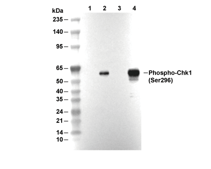 Lane 1: HeLa, Lane 2: HeLa (UV, 2 h), Lane 3: C6, Lane 4: C6 (UV, 2 h)