|
Come citare 1. Per la citazione nel testo (Materiali e Metodi): 2. Per la tabella delle risorse chiave: |
||
|
Numero verde: (877) 796-6397 -- Solo USA e Canada -- |
Fax: +1-832-582-8590 Ordini: +1-832-582-8158 |
Supporto tecnico: +1-832-582-8158 Ext:3 Si prega di fornire il numero dordine nelle-mail. Ci sforziamo di rispondere a tutte le richieste via e-mail entro un giorno lavorativo. |
Descrizione biologica
| Specificità | L'anticorpo Phospho-β Catenin (Ser37) [H12E11] rileva i livelli endogeni di β-catenina solo quando fosforilata alla serina 37. |
|---|---|
| Contesto | Phospho-β Catenin (Ser37) è una proteina multifunzionale appartenente alla famiglia delle armadillo, caratterizzata da 12 ripetizioni armadillo che formano una struttura superelicale con un solco carico positivamente che funge da interfaccia versatile per i partner di legame. È una proteina lunga 781 amminoacidi, composta dal dominio N-terminale (NTD), dodici domini armadillo (ARM) al centro della proteina e il dominio C-terminale (CTD). Svolge un duplice ruolo nell'adesione cellula-cellula mediata dalle caderine e nella regolazione trascrizionale mediata dalla segnalazione Wnt. Nell'adesione, la β-catenina stabilizza le caderine sulla membrana e recluta l'α-catenina per legarsi al citoscheletro di actina, mentre nella segnalazione agisce come co-attivatore nel complesso di trascrizione LEF/TCF in seguito all'attivazione di Wnt. La funzione della β-catenina è strettamente regolata dal suo stato di fosforilazione; la fosforilazione alla Ser37 (insieme a S33 e T41) da parte di GSK3β o PKC indirizza la β-catenina per l'ubiquitinazione e la degradazione proteasomica, prevenendo così una segnalazione Wnt aberrante. La fosforilazione del ligando può anche aumentare l'affinità di legame della β-catenina aumentando le cariche negative che interagiscono con il suo dominio armadillo. Le sue regioni N- e C-terminali non strutturate modulano ulteriormente la specificità e l'accessibilità dell'interazione. Nel complesso, l'adattabilità strutturale e la regolazione dipendente dalla fosforilazione della β-catenina sono fondamentali per i suoi ruoli dinamici nella morfogenesi tissutale, nelle decisioni sul destino cellulare e nel mantenimento dell'integrità tissutale. |
Informazioni sullutilizzo
| Applicazione | WB | Diluizione |
|
||
|---|---|---|---|---|---|
| Reattività | Human, Mouse, Rat | ||||
| Fonte | Rabbit Monoclonal Antibody | MW | 85 kDa | ||
| Tampone di conservazione | PBS, pH 7.2+50% Glycerol+0.05% BSA+0.01% NaN3 | Conservazione (Dalla data di ricevimento) |
-20°C (avoid freeze-thaw cycles), 2 years | ||
| WB |
Experimental Protocol:
Sample preparation
1. Tissue: Lyse the tissue sample by adding an appropriate volume of ice-cold RIPA/NP-40 Lysis Buffer (containing Protease Inhibitor Cocktail, Phosphatase Inhibitor Cocktail),and homogenize the tissue at a low temperature. 2. Adherent cell: Aspirate the culture medium and wash the cells with ice-cold PBS twice. Lyse the cells by adding an appropriate volume of RIPA/NP-40 Lysis Buffer (containing Protease Inhibitor Cocktail, Phosphatase Inhibitor Cocktail) and put the sample on ice for 5 min. 3. Suspension cell: Transfer the culture medium to a pre-cooled centrifuge tube. Centrifuge and aspirate the supernatant. Wash the cells with ice-cold PBS twice. Lyse the cells by adding an appropriate volume of RIPA/NP-40 Lysis Buffer (containing Protease Inhibitor Cocktail, Phosphatase Inhibitor Cocktail) and put the sample on ice for 5 min. 4. Place the lysate into a pre-cooled microcentrifuge tube. Centrifuge at 4°C for 15 min. Collect the supernatant;
5. Remove a small volume of lysate to determine the protein concentration;
6. Combine the lysate with protein loading buffer. Boil 20 µL sample under 95-100°C for 5 min. Centrifuge for 5 min after cool down on ice.
Electrophoretic separation
1. According to the concentration of extracted protein, load appropriate amount of protein sample and marker onto SDS-PAGE gels for electrophoresis. Recommended separating gel (lower gel) concentration: 10%. Reference Table for Selecting SDS-PAGE Separation Gel Concentrations 2. Power up 80V for 30 minutes. Then the power supply is adjusted (110 V~150 V), the Marker is observed, and the electrophoresis can be stopped when the indicator band of the predyed protein Marker where the protein is located is properly separated. (Note that the current should not be too large when electrophoresis, too large current (more than 150 mA) will cause the temperature to rise, affecting the result of running glue. If high currents cannot be avoided, an ice bath can be used to cool the bath.)
Transfer membrane
1. Take out the converter, soak the clip and consumables in the pre-cooled converter;
2. Activate PVDF membrane with methanol for 1 min and rinse with transfer buffer;
3. Install it in the order of "black edge of clip - sponge - filter paper - filter paper - glue -PVDF membrane - filter paper - filter paper - sponge - white edge of clip"; 4. The protein was electrotransferred to PVDF membrane. ( 0.45 µm PVDF membrane is recommended ) Reference Table for Selecting PVDF Membrane Pore Size Specifications Recommended conditions for wet transfer: 200 mA, 120 min. ( Note that the transfer conditions can be adjusted according to the protein size. For high-molecular-weight proteins, a higher current and longer transfer time are recommended. However, ensure that the transfer tank remains at a low temperature to prevent gel melting.)
Block
1. After electrotransfer, wash the film with TBST at room temperature for 5 minutes;
2. Incubate the film in the blocking solution ( recommending 5% BSA solution)
for 1 hour at room temperature;
3. Wash the film with TBST for 3 times, 5 minutes each time.
Antibody incubation
1. Use 5% skim milk powder to prepare the primary antibody working liquid (recommended dilution ratio for primary antibody 1:1000), gently shake and incubate with the film at 4°C overnight; 2. Wash the film with TBST 3 times, 5 minutes each time;
3. Add the secondary antibody to the blocking solution and incubate with the film gently at room temperature for 1 hour;
4. After incubation, wash the film with TBST 3 times for 5 minutes each time.
Antibody staining
1. Add the prepared ECL luminescent substrate (or select other color developing substrate according to the second antibody) and mix evenly;
2. Incubate with the film for 1 minute, remove excess substrate (keep the film moist), wrap with plastic film, and expose in the imaging system.
|
Riferimenti
|
Dati di applicazione
WB
Validato da Selleck
-
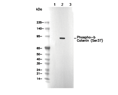 Lane 1: HEK-293, Lane 2: HEK-293 (Calyculin A, 50nM, 3 h), Lane 3: HEK-293 (Calyculin A, 50nM, 3 h; alkaline phosphatase, 1 h)