|
Come citare 1. Per la citazione nel testo (Materiali e Metodi): 2. Per la tabella delle risorse chiave: |
||
|
Numero verde: (877) 796-6397 -- Solo USA e Canada -- |
Fax: +1-832-582-8590 Ordini: +1-832-582-8158 |
Supporto tecnico: +1-832-582-8158 Ext:3 Si prega di fornire il numero dordine nelle-mail. Ci sforziamo di rispondere a tutte le richieste via e-mail entro un giorno lavorativo. |
Descrizione biologica
| Specificità | PFKFB2 Antibody [E19B24] rileva i livelli endogeni della proteina PFKFB2 totale. |
|---|---|
| Contesto | PFKFB2, l'enzima bifunzionale predominante nel cuore e nel muscolo 6-fosfofrutto-2-chinasi/fruttosio-2,6-bisfosfatasi, regola precisamente il flusso glicolitico controllando i livelli di fruttosio-2,6-bisfosfato, il più potente attivatore allosterico della fosfofrutto-1-chinasi, accelerando così la glicolisi e inibendo la gluconeogenesi, un ruolo particolarmente vitale nel metabolismo energetico cardiaco sotto stress, insulina e regolazione adrenergica. L'enzima forma un omodimero testa a testa, con ciascun monomero contenente un segmento regolatorio N-terminale, un dominio chinasico centrale con un'architettura bilobata simile all'adenilato chinasi che presenta un foglietto β a sei filamenti e α-eliche, e una tasca di legame ATP/fruttosio-6-fosfato con residui catalitici chiave che coordinano Mg²⁺-ATP. Il suo dominio fosfatasico C-terminale, omologo alle istidina fosfatasi, contiene anse flessibili che consentono l'accesso del fruttosio-2,6-bisfosfato per l'idrolisi tramite un relè di fosfoistidina, mentre la coda C-terminale ospita i siti Ser466/483 per la fosforilazione di Akt e PKA. L'attività chinasica sintetizza il fruttosio-2,6-bisfosfato dal fruttosio-6-fosfato e ATP tramite la chiusura del dominio che imita il movimento di esclusione dell'acqua dell'adenilato chinasi, consentendo un efficiente trasferimento fosforilico. Al contrario, l'attività bisfosfatasica idrolizza il fruttosio-2,6-bisfosfato nuovamente a fruttosio-6-fosfato tramite un attacco nucleofilo del motivo His-Pro-Pro che forma un intermedio fosfoenzimatico transitorio, e l'inibizione reciproca del substrato assicura una funzione di stato stazionario simile a un reostato. La fosforilazione dipendente da Akt a Ser483 durante la segnalazione PI3K/insulina aumenta l'attività chinasica per promuovere la glicolisi cardiaca dopo l'alimentazione, mentre la fosforilazione di PKA a Ser466/29 durante l'elevazione β-adrenergica e del cAMP inibisce la chinasi e attiva la fosfatasi, reindirizzando il metabolismo verso l'ossidazione dei grassi e fornendo protezione ischemica, mantenendo così un alto livello basale di fruttosio-2,6-bisfosfato per l'omeostasi dell'ATP. La disregolazione di PFKFB2, inclusa la commutazione di isoforma o la ridotta espressione, porta a cardiomiopatia diabetica con contrattilità compromessa, deficit glicolitici nell'insufficienza cardiaca e facilita la riprogrammazione metabolica di Warburg nel cancro. |
Informazioni sullutilizzo
| Applicazione | WB, IP, IF | Diluizione |
|
||||||
|---|---|---|---|---|---|---|---|---|---|
| Reattività | Human, Monkey | ||||||||
| Fonte | Rabbit Monoclonal Antibody | MW | 55 kDa | ||||||
| Tampone di conservazione | PBS, pH 7.2+50% Glycerol+0.05% BSA+0.01% NaN3 | Conservazione (Dalla data di ricevimento) |
-20°C (avoid freeze-thaw cycles), 2 years | ||||||
| WB |
Experimental Protocol:
Sample preparation
1. Tissue: Lyse the tissue sample by adding an appropriate volume of ice-cold RIPA/NP-40 Lysis Buffer (containing Protease Inhibitor Cocktail),and homogenize the tissue at a low temperature. 2. Adherent cell: Aspirate the culture medium and wash the cells with ice-cold PBS twice. Lyse the cells by adding an appropriate volume of RIPA/NP-40 Lysis Buffer (containing Protease Inhibitor Cocktail) and put the sample on ice for 5 min. 3. Suspension cell: Transfer the culture medium to a pre-cooled centrifuge tube. Centrifuge and aspirate the supernatant. Wash the cells with ice-cold PBS twice. Lyse the cells by adding an appropriate volume of RIPA/NP-40 Lysis Buffer (containing Protease Inhibitor Cocktail) and put the sample on ice for 5 min. 4. Place the lysate into a pre-cooled microcentrifuge tube. Centrifuge at 4°C for 15 min. Collect the supernatant;
5. Remove a small volume of lysate to determine the protein concentration;
6. Combine the lysate with protein loading buffer. Boil 20 µL sample under 95-100°C for 5 min. Centrifuge for 5 min after cool down on ice.
Electrophoretic separation
1. According to the concentration of extracted protein, load appropriate amount of protein sample and marker onto SDS-PAGE gels for electrophoresis. Recommended separating gel (lower gel) concentration: 10%. Reference Table for Selecting SDS-PAGE Separation Gel Concentrations 2. Power up 80V for 30 minutes. Then the power supply is adjusted (110 V~150 V), the Marker is observed, and the electrophoresis can be stopped when the indicator band of the predyed protein Marker where the protein is located is properly separated. (Note that the current should not be too large when electrophoresis, too large current (more than 150 mA) will cause the temperature to rise, affecting the result of running glue. If high currents cannot be avoided, an ice bath can be used to cool the bath.)
Transfer membrane
1. Take out the converter, soak the clip and consumables in the pre-cooled converter;
2. Activate PVDF membrane with methanol for 1 min and rinse with transfer buffer;
3. Install it in the order of "black edge of clip - sponge - filter paper - filter paper - glue -PVDF membrane - filter paper - filter paper - sponge - white edge of clip"; 4. The protein was electrotransferred to PVDF membrane. ( 0.45 µm PVDF membrane is recommended ) Reference Table for Selecting PVDF Membrane Pore Size Specifications Recommended conditions for wet transfer: 200 mA, 120 min. ( Note that the transfer conditions can be adjusted according to the protein size. For high-molecular-weight proteins, a higher current and longer transfer time are recommended. However, ensure that the transfer tank remains at a low temperature to prevent gel melting.)
Block
1. After electrotransfer, wash the film with TBST at room temperature for 5 minutes;
2. Incubate the film in the blocking solution for 1 hour at room temperature;
3. Wash the film with TBST for 3 times, 5 minutes each time.
Antibody incubation
1. Use 5% skim milk powder to prepare the primary antibody working liquid (recommended dilution ratio for primary antibody 1:1000), gently shake and incubate with the film at 4°C overnight; 2. Wash the film with TBST 3 times, 5 minutes each time;
3. Add the secondary antibody to the blocking solution and incubate with the film gently at room temperature for 1 hour;
4. After incubation, wash the film with TBST 3 times for 5 minutes each time.
Antibody staining
1. Add the prepared ECL luminescent substrate (or select other color developing substrate according to the second antibody) and mix evenly;
2. Incubate with the film for 1 minute, remove excess substrate (keep the film moist), wrap with plastic film, and expose in the imaging system.
|
| IF |
Experimental Protocol:
Sample Preparation
1. Adherent Cells: Place a clean, sterile coverslip in a culture dish. Once the cells grow to near confluence as a monolayer, remove the coverslip for further use.
2. Suspension Cells: Seed the cells onto a clean, sterile slide coated with poly-L-lysine.
3. Frozen Sections: Allow the slide to thaw at room temperature. Wash it with pure water or PBS for 2 times, 3 minutes each time.
4. Paraffin Sections: Deparaffinization and rehydration. Wash the slide with pure water or PBS for 3 times, 3 minutes each time. Then perform antigen retrieval.
Fixation
1. Fix the cell coverslips/spots or tissue sections at room temperature using a fixative such as 4% paraformaldehyde (4% PFA) for 10-15 minutes.
2. Wash the sample with PBS for 3 times, 3 minutes each time.
Permeabilization
1.Add a detergent such as 0.1–0.3% Triton X-100 to the sample and incubate at room temperature for 10–20 minutes.
(Note: This step is only required for intracellular antigens. For antigens expressed on the cell membrane, this step is unnecessary.)
Wash the sample with PBS for 3 times, 3 minutes each time.
Blocking
Add blocking solution and incubate at room temperature for at least 1 hour. (Common blocking solutions include: serum from the same source as the secondary antibody, BSA, or goat serum.)
Note: Ensure the sample remains moist during and after the blocking step to prevent drying, which can lead to high background.
Immunofluorescence Staining (Day 1)
1. Remove the blocking solution and add the diluted primary antibody.
2. Incubate the sample in a humidified chamber at 4°C overnight.
Immunofluorescence Staining (Day 2)
1. Remove the primary antibody and wash with PBST for 3 times, 5 minutes each time.
2. Add the diluted fluorescent secondary antibody and incubate in the dark at 4°C for 1–2 hours.
3. Remove the secondary antibody and wash with PBST for 3 times, 5 minutes each time.
4. Add diluted DAPI and incubate at room temperature in the dark for 5–10 minutes.
5. Wash with PBST for 3 times, 5 minutes each time.
Mounting
1. Mount the sample with an anti-fade mounting medium.
2. Allow the slide to dry at room temperature overnight in the dark.
3. Store the slide in a slide storage box at 4°C, protected from light.
|
Riferimenti
|
Dati di applicazione
WB
Validato da Selleck
-
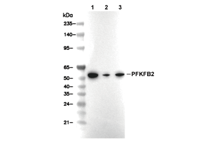 Lane 1: LNCAP, Lane 2: HepG2, Lane 3: MCF7