Dati tecnici
| Formula | C13H13N3OS |
||||||
| Peso molecolare | 259.33 | Numero CAS | 4311-88-0 | ||||
| Solubilità (25°C)* | In vitro | DMSO | 52 mg/mL (200.51 mM) | ||||
| Water | Insoluble | ||||||
| Ethanol | Insoluble | ||||||
| In vivo (Aggiungere i solventi al prodotto singolarmente e in ordine.) |
|
||||||
|
* <1 mg/ml significa leggermente solubile o insolubile. * Si prega di notare che Selleck testa la solubilità di tutti i composti internamente e la solubilità effettiva può differire leggermente dai valori pubblicati. Ciò è normale ed è dovuto a leggere variazioni da lotto a lotto. * Spedizione a temperatura ambiente (I test di stabilità mostrano che questo prodotto può essere spedito senza alcuna misura di raffreddamento.) |
|||||||
Preparazione delle soluzioni stock
Attività biologica
| Descrizione | Necrostatin-1 (Nec-1) è un inibitore specifico di RIP1 (RIPK1) e inibisce la necroptosi indotta da TNF-α con un EC50 di 490 nM in cellule 293T. Necrostatin-1 blocca anche IDO e sopprime l'Autophagy e l'Apoptosis. | ||||
|---|---|---|---|---|---|
| Target |
|
||||
| In vitro | La Necrostatin-1 (1-100 μM) inibisce l'autofosforilazione di RIP1 sovraespresso ed endogeno. Si è riscontrato che RIP1 è il bersaglio cellulare primario responsabile dell'attività antinecroptotica di questo composto. Questa sostanza chimica sopprime efficacemente la morte cellulare necroptotica innescata da una serie di stimoli in una varietà di tipi cellulari. Essa, precedentemente identificata come inibitore a piccole molecole della necroptosi, inibisce la necroptosi indotta dalla RIP kinase e inibisce la necroptosi indotta da TNF-α nelle cellule Jurkat con una EC50 di 490 nM. |
||||
| In vivo | Necrostatin-1 (Nec-1) è un inibitore specifico a piccole molecole della proteina chinasi 1 (RIPK1) interagente con il recettore che inibisce specificamente la fosforilazione di questo composto. |
||||
| Caratteristiche | Uno strumento potente per caratterizzare il ruolo della necroptosi con un bersaglio primario caratterizzato. |
Protocollo (da riferimento)
| Saggio della chinasi: |
|
|---|---|
| Saggio cellulare: |
|
| Studio sugli animali: |
|
Riferimenti
|
Convalida del prodotto da parte del cliente
-
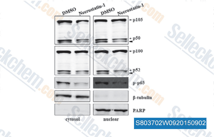
Dati da [ , , J Cell Mol Med, 2015, 19(5): 1042-54 ]
-
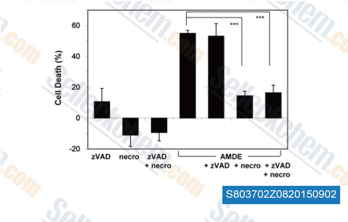
Dati da [ , , PLoS One, 2015, 10(3): e0122083 ]
-
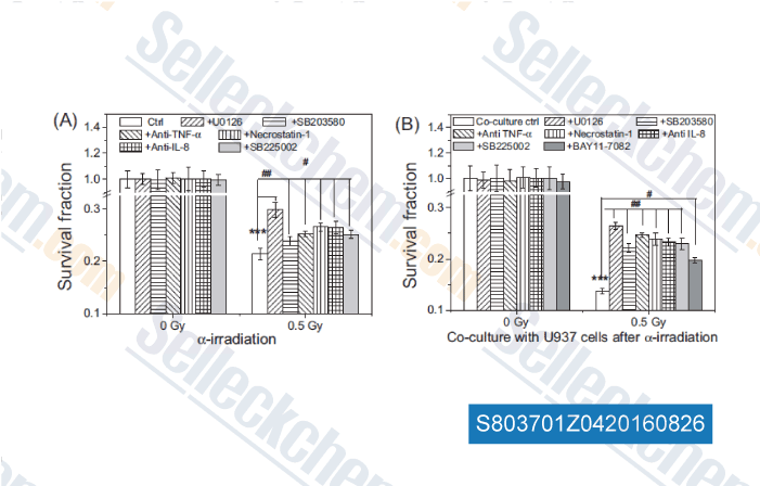
Dati da [ , , Mutation Research, 2016, 789:1-8. ]
Sellecks Necrostatin-1 (Nec-1) È stato citato da 321 Pubblicazioni
| Soluble tissue factor generated by necroptosis-triggered shedding is responsible for thrombosis [ Cell Res, 2025, 10.1038/s41422-025-01167-8] | PubMed: 40940518 |
| The noncanonical function of liver-type phosphofructokinase potentiates the efficacy of HDAC inhibitors in cancer [ Signal Transduct Target Ther, 2025, 10(1):341] | PubMed: 41083431 |
| LINE-1 ORF1p Mimics Viral Innate Immune Evasion Mechanisms in Pancreatic Ductal Adenocarcinoma [ Cancer Discov, 2025, 10.1158/2159-8290.CD-24-1317] | PubMed: 39919290 |
| Ferroptosis-activating metabolite acrolein antagonizes necroptosis and anti-cancer therapeutics [ Nat Commun, 2025, 16(1):4919] | PubMed: 40425585 |
| Harnessing the FGFR2/NF2/YAP signaling-dependent necroptosis to develop an FGFR2/IL-8 dual blockade therapeutic strategy [ Nat Commun, 2025, 16(1):4128] | PubMed: 40319089 |
| Targeting pancreatic cancer glutamine dependency confers vulnerability to GPX4-dependent ferroptosis [ Cell Rep Med, 2025, 6(2):101928] | PubMed: 39879992 |
| GCLC desuccinylation regulated by oxidative stress protects human cancer cells from ferroptosis [ Cell Death Differ, 2025, 32(9):1679-1690] | PubMed: 40188196 |
| Carbon ion combined photon radiotherapy induces ferroptosis via NCOA4-mediated ferritinophagy in glioblastoma [ Redox Biol, 2025, 86:103865] | PubMed: 40925125 |
| SLC25A1 and ACLY maintain cytosolic acetyl-CoA and regulate ferroptosis susceptibility via FSP1 acetylation [ EMBO J, 2025, 10.1038/s44318-025-00369-5] | PubMed: 39881208 |
| Akt isoform specificity drives intrinsic immune regulation during HSV-1 infection [ Proc Natl Acad Sci U S A, 2025, 122(27):e2504962122] | PubMed: 40601626 |
POLITICA DI RESO
La politica di reso incondizionato di Selleck Chemical garantisce ai nostri clienti unesperienza di acquisto online senza problemi. Se non sei in alcun modo soddisfatto del tuo acquisto, puoi restituire qualsiasi articolo entro 7 giorni dalla ricezione. In caso di problemi di qualità del prodotto, sia relativi al protocollo che al prodotto, puoi restituire qualsiasi articolo entro 365 giorni dalla data di acquisto originale. Si prega di seguire le istruzioni seguenti per la restituzione dei prodotti.
SPEDIZIONE E CONSERVAZIONE
I prodotti Selleck vengono trasportati a temperatura ambiente. Se ricevi il prodotto a temperatura ambiente, ti assicuriamo che il Dipartimento di Ispezione Qualità di Selleck ha condotto esperimenti per verificare che la conservazione a temperatura normale per un mese non influirà sullattività biologica dei prodotti in polvere. Dopo la raccolta, conservare il prodotto secondo le indicazioni descritte nella scheda tecnica. La maggior parte dei prodotti Selleck è stabile nelle condizioni raccomandate.
NON PER USO UMANO, DIAGNOSTICO VETERINARIO O TERAPEUTICO.