|
Come citare 1. Per la citazione nel testo (Materiali e Metodi): 2. Per la tabella delle risorse chiave: |
||
|
Numero verde: (877) 796-6397 -- Solo USA e Canada -- |
Fax: +1-832-582-8590 Ordini: +1-832-582-8158 |
Supporto tecnico: +1-832-582-8158 Ext:3 Si prega di fornire il numero dordine nelle-mail. Ci sforziamo di rispondere a tutte le richieste via e-mail entro un giorno lavorativo. |
Descrizione biologica
| Specificità | MCP1 Antibody [M3B19] rileva i livelli endogeni della proteina MCP1 totale. |
|---|---|
| Contesto | La proteina chemiotattica dei monociti-1 (MCP-1/CCL2) è un membro della famiglia delle chemochine C-C e funziona come un potente fattore chemiotattico per i monociti. È considerata identica a JE, un gene originariamente identificato nei fibroblasti di topo come indotto dal fattore di crescita derivato dalle piastrine. Il gene umano MCP-1 si trova sul cromosoma 17q11.2 e codifica una proteina di 76 aminoacidi con un peso molecolare di circa 13 kDa. MCP-1 appartiene a una sottofamiglia di chemochine che include almeno quattro membri: MCP-1, MCP-2, MCP-3 e MCP-4. CCL2 è prodotto da una vasta gamma di tipi cellulari, sia costitutivamente che in risposta a stimoli come stress ossidativo, citochine e fattori di crescita. Le sue fonti includono cellule endoteliali, fibroblasti, cellule epiteliali, cellule muscolari lisce, cellule mesangiali, astrociti, monociti e microglia, tipi cellulari che svolgono ruoli importanti nella difesa immunitaria antivirale sia nella circolazione che nei tessuti. Funzionalmente, CCL2 dirige la migrazione e l'infiltrazione di monociti, cellule T della memoria e cellule natural killer (NK). Gli effetti biologici di CCL2 sono mediati attraverso il suo recettore, CCR2, la cui espressione è più ristretta rispetto a quella di CCL2. CCR2 esiste in due isoforme a splicing alternativo, CCR2A e CCR2B, che differiscono solo nelle loro regioni C-terminali. |
Informazioni sullutilizzo
| Applicazione | IHC | Diluizione |
|
|---|---|---|---|
| Reattività | Mouse | ||
| Fonte | Rat Monoclonal Antibody | MW | 11 kDa |
| Tampone di conservazione | PBS, pH 7.2+50% Glycerol+0.05% BSA+0.01% NaN3 | Conservazione (Dalla data di ricevimento) |
-20°C (avoid freeze-thaw cycles), 2 years |
| IHC |
Experimental Protocol:
Deparaffinization/Rehydration
1. Deparaffinize/hydrate sections:
2. Incubate sections in three washes of xylene for 5 min each.
3. Incubate sections in two washes of 100% ethanol for 10 min each.
4. Incubate sections in two washes of 95% ethanol for 10 min each.
5. Wash sections two times in dH2O for 5 min each.
6.Antigen retrieval: For Citrate: Heat slides in a microwave submersed in 1X citrate unmasking solution until boiling is initiated; continue with 10 min at a sub-boiling temperature (95°-98°C). Cool slides on bench top for 30 min.
Staining
1. Wash sections in dH2O three times for 5 min each.
2. Incubate sections in 3% hydrogen peroxide for 10 min.
3. Wash sections in dH2O two times for 5 min each.
4. Wash sections in wash buffer for 5 min.
5. Block each section with 100–400 µl of blocking solution for 1 hr at room temperature.
6. Remove blocking solution and add 100–400 µl primary antibody diluent in to each section. Incubate overnight at 4°C.
7. Remove antibody solution and wash sections with wash buffer three times for 5 min each.
8. Cover section with 1–3 drops HRPas needed. Incubate in a humidified chamber for 30 min at room temperature.
9. Wash sections three times with wash buffer for 5 min each.
10. Add DAB Chromogen Concentrate to DAB Diluent and mix well before use.
11. Apply 100–400 µl DAB to each section and monitor closely. 1–10 min generally provides an acceptable staining intensity.
12. Immerse slides in dH2O.
13. If desired, counterstain sections with hematoxylin.
14. Wash sections in dH2O two times for 5 min each.
15. Dehydrate sections: Incubate sections in 95% ethanol two times for 10 sec each; Repeat in 100% ethanol, incubating sections two times for 10 sec each; Repeat in xylene, incubating sections two times for 10 sec each.
16. Mount sections with coverslips and mounting medium.
|
Riferimenti
|
Dati di applicazione
IHC
Validato da Selleck
-
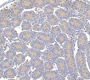 Immunohistochemical analysis of formalin fixed paraffin embedded mouse testicles tissue with F3225 at 1:10 dilution.