|
Come citare 1. Per la citazione nel testo (Materiali e Metodi): 2. Per la tabella delle risorse chiave: |
||
|
Numero verde: (877) 796-6397 -- Solo USA e Canada -- |
Fax: +1-832-582-8590 Ordini: +1-832-582-8158 |
Supporto tecnico: +1-832-582-8158 Ext:3 Si prega di fornire il numero dordine nelle-mail. Ci sforziamo di rispondere a tutte le richieste via e-mail entro un giorno lavorativo. |
Descrizione biologica
| Specificità | Influenza A virus (H5N1/HA1) Antibody [L10F5] rileva livelli esogeni della proteina totale Influenza A virus (H5N1/HA1). |
|---|---|
| Contesto | Influenza A virus (H5N1/HA1) è la subunità N-terminale legante il recettore dell'emoagglutinina (HA), una glicoproteina di fusione virale di classe I sintetizzata come precursore HA0 e scissa dalla furina dell'ospite in siti multibasici (caratteristici dell'H5N1 altamente patogeno) in HA1 (residui ~1-328) e HA2 (~221 aa) legati da ponti disolfuro. HA1 forma un dominio di testa globulare distale alla membrana sulla stelo di HA2, composto da foglietti β antiparalleli con motivi α/β misti, caratterizzato dal sito di legame del recettore dell'acido sialico (RBS, residui chiave Gln226, Gly228 per la preferenza di legame α2,3), dalle anse 130/220/elica 190 che definiscono i siti antigenici e dai glicani N-legati (~7 siti) che modulano l'immunogenicità e la stabilità. L'HA1 trimerico media l'attaccamento ai glicani sialilati sugli epiteli respiratori dell'ospite, innescando l'endocitosi mediata dal recettore; l'acidificazione endosomale (pH 5,0-5,5) induce la dissociazione irreversibile di HA1 da HA2, esponendo il peptide di fusione di HA2 per la fusione della membrana virale-endosomale e il rilascio di vRNP nel citoplasma. La maggiore stabilità acida e la scissione multibasica dell'HA1 dell'H5N1 consentono la replicazione sistemica, l'elevata virulenza (60% di CFR umano) e il potenziale zoonotico tramite adattamenti del legame al recettore (es. Q226L). |
Informazioni sullutilizzo
| Applicazione | WB, ELISA | Diluizione |
|
||
|---|---|---|---|---|---|
| Reattività | Influenza A | ||||
| Fonte | Mouse Monoclonal Antibody | MW | 64 kDa | ||
| Tampone di conservazione | PBS, pH 7.2+50% Glycerol+0.05% BSA+0.01% NaN3 | Conservazione (Dalla data di ricevimento) |
-20°C (avoid freeze-thaw cycles), 2 years | ||
| WB |
Experimental Protocol:
Sample preparation
1. Tissue: Lyse the tissue sample by adding an appropriate volume of ice-cold RIPA/NP-40 Lysis Buffer (containing Protease Inhibitor Cocktail),and homogenize the tissue at a low temperature. 2. Adherent cell: Aspirate the culture medium and wash the cells with ice-cold PBS twice. Lyse the cells by adding an appropriate volume of RIPA/NP-40 Lysis Buffer (containing Protease Inhibitor Cocktail) and put the sample on ice for 5 min. 3. Suspension cell: Transfer the culture medium to a pre-cooled centrifuge tube. Centrifuge and aspirate the supernatant. Wash the cells with ice-cold PBS twice. Lyse the cells by adding an appropriate volume of RIPA/NP-40 Lysis Buffer (containing Protease Inhibitor Cocktail) and put the sample on ice for 5 min. 4. Place the lysate into a pre-cooled microcentrifuge tube. Centrifuge at 4°C for 15 min. Collect the supernatant;
5. Remove a small volume of lysate to determine the protein concentration;
6. Combine the lysate with protein loading buffer. Boil 20 µL sample under 95-100°C for 5 min. Centrifuge for 5 min after cool down on ice.
Electrophoretic separation
1. According to the concentration of extracted protein, load appropriate amount of protein sample and marker onto SDS-PAGE gels for electrophoresis. Recommended separating gel (lower gel) concentration: 10%. Reference Table for Selecting SDS-PAGE Separation Gel Concentrations 2. Power up 80V for 30 minutes. Then the power supply is adjusted (110 V~150 V), the Marker is observed, and the electrophoresis can be stopped when the indicator band of the predyed protein Marker where the protein is located is properly separated. (Note that the current should not be too large when electrophoresis, too large current (more than 150 mA) will cause the temperature to rise, affecting the result of running glue. If high currents cannot be avoided, an ice bath can be used to cool the bath.)
Transfer membrane
1. Take out the converter, soak the clip and consumables in the pre-cooled converter;
2. Activate PVDF membrane with methanol for 1 min and rinse with transfer buffer;
3. Install it in the order of "black edge of clip - sponge - filter paper - filter paper - glue -PVDF membrane - filter paper - filter paper - sponge - white edge of clip"; 4. The protein was electrotransferred to PVDF membrane. ( 0.45 µm PVDF membrane is recommended ) Reference Table for Selecting PVDF Membrane Pore Size Specifications Recommended conditions for wet transfer: 200 mA, 120 min. ( Note that the transfer conditions can be adjusted according to the protein size. For high-molecular-weight proteins, a higher current and longer transfer time are recommended. However, ensure that the transfer tank remains at a low temperature to prevent gel melting.)
Block
1. After electrotransfer, wash the film with TBST at room temperature for 5 minutes;
2. Incubate the film in the blocking solution for 1 hour at room temperature;
3. Wash the film with TBST for 3 times, 5 minutes each time.
Antibody incubation
1. Use 5% skim milk powder to prepare the primary antibody working liquid (recommended dilution ratio for primary antibody 1:3000), gently shake and incubate with the film at 4°C overnight; 2. Wash the film with TBST 3 times, 5 minutes each time;
3. Add the secondary antibody to the blocking solution and incubate with the film gently at room temperature for 1 hour;
4. After incubation, wash the film with TBST 3 times for 5 minutes each time.
Antibody staining
1. Add the prepared ECL luminescent substrate (or select other color developing substrate according to the second antibody) and mix evenly;
2. Incubate with the film for 1 minute, remove excess substrate (keep the film moist), wrap with plastic film, and expose in the imaging system.
|
Riferimenti
|
Dati di applicazione
WB
Validato da Selleck
-
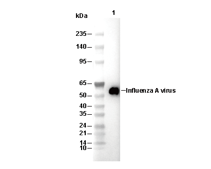 Lane 1: H5N1 recombinant protein