|
Come citare 1. Per la citazione nel testo (Materiali e Metodi): 2. Per la tabella delle risorse chiave: |
||
|
Numero verde: (877) 796-6397 -- Solo USA e Canada -- |
Fax: +1-832-582-8590 Ordini: +1-832-582-8158 |
Supporto tecnico: +1-832-582-8158 Ext:3 Si prega di fornire il numero dordine nelle-mail. Ci sforziamo di rispondere a tutte le richieste via e-mail entro un giorno lavorativo. |
Descrizione biologica
| Specificità | HACE1 Antibody [A17B18] riconosce i livelli endogeni della proteina HACE1 totale. |
|---|---|
| Contesto | Il gene HACE1 (HECT domain and ankyrin repeat-containing E3 ubiquitin-protein ligase 1) si trova sul cromosoma 6 e codifica una proteina composta da 909 amminoacidi. HACE1 è fortemente espresso in vari tessuti umani, inclusi cuore, cervello, placenta, pancreas e reni sia fetali che adulti. L'mRNA di HACE1 è espresso ubiquitariamente nei tessuti umani normali. Livelli ridotti di espressione di HACE1 sono fortemente associati all'ipermetilazione di due isole CpG situate a monte del suo locus genico, suggerendo che la regolazione epigenetica giochi un ruolo nel suo silenziamento. HACE1 è frequentemente sottoregolato in diversi tipi di cancro, inclusi linfoma a cellule T/natural killer (NKTCL), cancro colorettale e cancro gastrico. A livello subcellulare, HACE1 si localizza principalmente nel reticolo endoplasmatico e nel citosol, sebbene tipicamente vengano rilevati solo bassi livelli di proteina endogena. Funzionalmente, HACE1 agisce come una E3 Ligase e collabora con l'enzima E2 UBCH7 per catalizzare l'ubiquitinazione delle proteine bersaglio. È notevolmente coinvolto nella degradazione dipendente dalla fosforilazione della ciclina D1, giocando così un ruolo nell'inibire la progressione del ciclo cellulare. HACE1 si lega anche preferenzialmente alla forma attiva, legata al GTP, della piccola GTPasi Rac1 e promuove la sua poliubiquitinazione. Questa attività è essenziale per la degradazione di Rac1 in risposta al fattore necrotizzante citotossico 1 (CNF1), facilitando l'invasione batterica delle monostrati di cellule endoteliali e evidenziando un ruolo per HACE1 nei meccanismi di difesa immunitaria innata. La perdita di HACE1 porta allo sviluppo spontaneo di tumori a insorgenza tardiva, supportando ulteriormente la sua funzione di soppressore tumorale. Il gene si trova all'interno della regione cromosomica 6q21, che è un hotspot implicato in molteplici tumori umani, sottolineando la sua importanza clinica nella tumorigenesi. |
Informazioni sullutilizzo
| Applicazione | WB | Diluizione |
|
||
|---|---|---|---|---|---|
| Reattività | Mouse, Rat, Human | ||||
| Fonte | Rabbit Monoclonal Antibody | MW | 102 kDa | ||
| Tampone di conservazione | PBS, pH 7.2+50% Glycerol+0.05% BSA+0.01% NaN3 | Conservazione (Dalla data di ricevimento) |
-20°C (avoid freeze-thaw cycles), 2 years | ||
| WB |
Experimental Protocol:
Sample preparation
1. Tissue: Lyse the tissue sample by adding an appropriate volume of ice-cold RIPA/NP-40 Lysis Buffer (containing Protease Inhibitor Cocktail),and homogenize the tissue at a low temperature. 2. Adherent cell: Aspirate the culture medium and wash the cells with ice-cold PBS twice. Lyse the cells by adding an appropriate volume of RIPA/NP-40 Lysis Buffer (containing Protease Inhibitor Cocktail) and put the sample on ice for 5 min. 3. Suspension cell: Transfer the culture medium to a pre-cooled centrifuge tube. Centrifuge and aspirate the supernatant. Wash the cells with ice-cold PBS twice. Lyse the cells by adding an appropriate volume of RIPA/NP-40 Lysis Buffer (containing Protease Inhibitor Cocktail) and put the sample on ice for 5 min. 4. Place the lysate into a pre-cooled microcentrifuge tube. Centrifuge at 4°C for 15 min. Collect the supernatant;
5. Remove a small volume of lysate to determine the protein concentration;
6. Combine the lysate with protein loading buffer. Boil 20 µL sample under 95-100°C for 5 min. Centrifuge for 5 min after cool down on ice.
Electrophoretic separation
1. According to the concentration of extracted protein, load appropriate amount of protein sample and marker onto SDS-PAGE gels for electrophoresis. Recommended separating gel (lower gel) concentration: 5%. Reference Table for Selecting SDS-PAGE Separation Gel Concentrations 2. Power up 80V for 30 minutes. Then the power supply is adjusted (110 V~150 V), the Marker is observed, and the electrophoresis can be stopped when the indicator band of the predyed protein Marker where the protein is located is properly separated. (Note that the current should not be too large when electrophoresis, too large current (more than 150 mA) will cause the temperature to rise, affecting the result of running glue. If high currents cannot be avoided, an ice bath can be used to cool the bath.)
Transfer membrane
1. Take out the converter, soak the clip and consumables in the pre-cooled converter;
2. Activate PVDF membrane with methanol for 1 min and rinse with transfer buffer;
3. Install it in the order of "black edge of clip - sponge - filter paper - filter paper - glue -PVDF membrane - filter paper - filter paper - sponge - white edge of clip"; 4. The protein was electrotransferred to PVDF membrane. ( 0.45 µm PVDF membrane is recommended ) Reference Table for Selecting PVDF Membrane Pore Size Specifications Recommended conditions for wet transfer: 200 mA, 120 min. ( Note that the transfer conditions can be adjusted according to the protein size. For high-molecular-weight proteins, a higher current and longer transfer time are recommended. However, ensure that the transfer tank remains at a low temperature to prevent gel melting.)
Block
1. After electrotransfer, wash the film with TBST at room temperature for 5 minutes;
2. Incubate the film in the blocking solution for 1 hour at room temperature;
3. Wash the film with TBST for 3 times, 5 minutes each time.
Antibody incubation
1. Use 5% skim milk powder to prepare the primary antibody working liquid (recommended dilution ratio for primary antibody 1:1000), gently shake and incubate with the film at 4°C overnight; 2. Wash the film with TBST 3 times, 5 minutes each time;
3. Add the secondary antibody to the blocking solution and incubate with the film gently at room temperature for 1 hour;
4. After incubation, wash the film with TBST 3 times for 5 minutes each time.
Antibody staining
1. Add the prepared ECL luminescent substrate (or select other color developing substrate according to the second antibody) and mix evenly;
2. Incubate with the film for 1 minute, remove excess substrate (keep the film moist), wrap with plastic film, and expose in the imaging system.
|
Riferimenti
|
Dati di applicazione
WB
Validato da Selleck
-
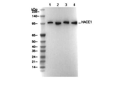 Lane 1: HEK293, Lane 2: SH-SY5Y, Lane 3: Mouse brain, Lane 4: Rat brain