|
Come citare 1. Per la citazione nel testo (Materiali e Metodi): 2. Per la tabella delle risorse chiave: |
||
|
Numero verde: (877) 796-6397 -- Solo USA e Canada -- |
Fax: +1-832-582-8590 Ordini: +1-832-582-8158 |
Supporto tecnico: +1-832-582-8158 Ext:3 Si prega di fornire il numero dordine nelle-mail. Ci sforziamo di rispondere a tutte le richieste via e-mail entro un giorno lavorativo. |
Descrizione biologica
| Specificità | Glutathione Antibody [A11G7] rileva i livelli endogeni della proteina totale Glutathione. |
|---|---|
| Contesto | Il Glutathione (GSH) è un antiossidante tiolico tripeptidico a basso peso molecolare composto da glutammato, cisteina e glicina, sintetizzato tramite due passaggi sequenziali ATP-dipendenti catalizzati dalla glutammato-cisteina ligasi e dalla glutatione sintetasi. Esiste principalmente in forme ridotta (GSH) e disolfuro ossidata (GSSG), che si interconvertono per regolare l'equilibrio redox cellulare. Il GSH presenta un legame peptidico γ-glutamilico unico tra il γ-carbossile del glutammato e il gruppo amminico della cisteina, conferendo resistenza alla degradazione da parte della γ-glutammil ciclotrasferasi e della maggior parte delle peptidasi; il tiolo della cisteina (-SH) agisce come centro nucleofilo per la chimica redox, mentre la glicina stabilizza il C-terminale. Il GSH scansiona direttamente le specie reattive dell'ossigeno e dell'azoto (ROS/RNS) tramite scambio tiolo-disolfuro, serve come riducente essenziale per le glutatione perossidasi (GPx) che detossificano i perossidi (con il GSSG ridotto nuovamente a GSH dalla glutatione reduttasi utilizzando NADPH), e coniuga xenobiotici elettrofili, metalli pesanti e tossine endogene attraverso le glutatione S-transferasi (GSTs) per la detossificazione di fase II e l'esportazione di acido mercapturico. Questi processi mantengono l'omeostasi dei solfidrili proteici, supportano l'espressione genica antiossidante guidata da Nrf2, facilitano l'assemblaggio dei cluster ferro-zolfo e il traffico dei metalli, e modulano le vie di segnalazione come NF-κB. L'esaurimento del GSH è implicato in malattie correlate allo stress ossidativo, tra cui il morbo di Parkinson, la cirrosi epatica, il cancro, il diabete e l'invecchiamento precoce. |
Informazioni sullutilizzo
| Applicazione | IF, FCM | Diluizione |
|
||||
|---|---|---|---|---|---|---|---|
| Reattività | |||||||
| Fonte | Mouse Monoclonal Antibody | MW | |||||
| Tampone di conservazione | PBS, pH 7.2+50% Glycerol+0.05% BSA+0.01% NaN3 | Conservazione (Dalla data di ricevimento) |
-20°C (avoid freeze-thaw cycles), 2 years | ||||
| IF |
Experimental Protocol:
Sample Preparation
1. Adherent Cells: Place a clean, sterile coverslip in a culture dish. Once the cells grow to near confluence as a monolayer, remove the coverslip for further use.
2. Suspension Cells: Seed the cells onto a clean, sterile slide coated with poly-L-lysine.
3. Frozen Sections: Allow the slide to thaw at room temperature. Wash it with pure water or PBS for 2 times, 3 minutes each time.
4. Paraffin Sections: Deparaffinization and rehydration. Wash the slide with pure water or PBS for 3 times, 3 minutes each time. Then perform antigen retrieval.
Fixation
1. Fix the cell coverslips/spots or tissue sections at room temperature using a fixative such as 4% paraformaldehyde (4% PFA) for 10-15 minutes.
2. Wash the sample with PBS for 3 times, 3 minutes each time.
Permeabilization
1.Add a detergent such as 0.1–0.3% Triton X-100 to the sample and incubate at room temperature for 10–20 minutes.
(Note: This step is only required for intracellular antigens. For antigens expressed on the cell membrane, this step is unnecessary.)
Wash the sample with PBS for 3 times, 3 minutes each time.
Blocking
Add blocking solution and incubate at room temperature for at least 1 hour. (Common blocking solutions include: serum from the same source as the secondary antibody, BSA, or goat serum.)
Note: Ensure the sample remains moist during and after the blocking step to prevent drying, which can lead to high background.
Immunofluorescence Staining (Day 1)
1. Remove the blocking solution and add the diluted primary antibody.
2. Incubate the sample in a humidified chamber at 4°C overnight.
Immunofluorescence Staining (Day 2)
1. Remove the primary antibody and wash with PBST for 3 times, 5 minutes each time.
2. Add the diluted fluorescent secondary antibody and incubate in the dark at 4°C for 1–2 hours.
3. Remove the secondary antibody and wash with PBST for 3 times, 5 minutes each time.
4. Add diluted DAPI and incubate at room temperature in the dark for 5–10 minutes.
5. Wash with PBST for 3 times, 5 minutes each time.
Mounting
1. Mount the sample with an anti-fade mounting medium.
2. Allow the slide to dry at room temperature overnight in the dark.
3. Store the slide in a slide storage box at 4°C, protected from light.
|
Riferimenti
|
Dati di applicazione
IF
Validato da Selleck
-
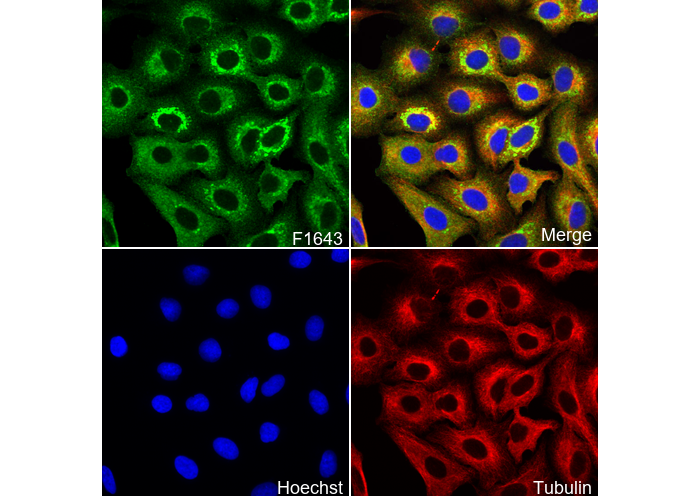 Immunofluorescent analysis of A549 cells using F1643 (green, 1:100), Hoechst (blue) and tubulin (Red).