|
Come citare 1. Per la citazione nel testo (Materiali e Metodi): 2. Per la tabella delle risorse chiave: |
||
|
Numero verde: (877) 796-6397 -- Solo USA e Canada -- |
Fax: +1-832-582-8590 Ordini: +1-832-582-8158 |
Supporto tecnico: +1-832-582-8158 Ext:3 Si prega di fornire il numero dordine nelle-mail. Ci sforziamo di rispondere a tutte le richieste via e-mail entro un giorno lavorativo. |
Descrizione biologica
| Specificità | GIV Antibody [K1N14] riconosce i livelli endogeni della proteina GIV totale. |
|---|---|
| Contesto | GIV (modulatore di segnalazione della proteina G) è un fattore di scambio di nucleotidi guaninici (GEF) non recettoriale critico che modula la segnalazione del recettore accoppiato a proteine G (GPCR) attivando le subunità Gαi. Comprende un dominio N-terminale che interagisce con il recettore del fattore di crescita epidermico (EGFR) e un dominio GEF C-terminale che attiva Gαi, influenzando così vari processi cellulari. GIV svolge un ruolo vitale nella regolazione dell'equilibrio tra migrazione e proliferazione cellulare, due processi essenziali per l'invasione tumorale e la metastasi. Dopo l'attivazione da parte dei fattori di crescita, GIV promuove segnali motogenici tramite PI3K e PLCγ1, facilitando il movimento cellulare, mentre sopprime i segnali mitogenici come ERK e STAT5b che sono coinvolti nella divisione cellulare. Nelle cellule tumorali, lo splicing alternativo può produrre una variante deficiente di GEF, GIVΔCT, che interrompe l'attivazione di Gαi, portando ad un aumento della proliferazione cellulare e una riduzione della migrazione, tratti caratteristici delle cellule tumorali meno invasive. Questa disregolazione dello splicing è particolarmente evidente nei tumori, dove l'espressione di GIV differisce tra le fasi. Il GIV a lunghezza intera (GIV-fl) è più prevalente nelle cellule tumorali invasive, mentre la sua variante spliced alternativamente si trova in quelle scarsamente invasive. Inoltre, GIV influenza il traffico e la degradazione dell'EGFR, regolando così il turnover del recettore e le dinamiche di segnalazione. |
Informazioni sullutilizzo
| Applicazione | WB, IP, IF, FCM | Diluizione |
|
||||||||
|---|---|---|---|---|---|---|---|---|---|---|---|
| Reattività | Human, Mouse, Rat | ||||||||||
| Fonte | Rabbit Monoclonal Antibody | MW | 132 kDa, 216 kDa, 69 kDa | ||||||||
| Tampone di conservazione | PBS, pH 7.2+50% Glycerol+0.05% BSA+0.01% NaN3 | Conservazione (Dalla data di ricevimento) |
-20°C (avoid freeze-thaw cycles), 2 years | ||||||||
| WB |
Experimental Protocol:
Sample preparation
1. Tissue: Lyse the tissue sample by adding an appropriate volume of ice-cold RIPA/NP-40 Lysis Buffer (containing Protease Inhibitor Cocktail),and homogenize the tissue at a low temperature or lyse it by sonication on ice, then incubate on ice for 30 minutes. 2. Adherent cell: Aspirate the culture medium and transfer the cells into an EP tube. Wash the cells with ice-cold PBS twice. Add an appropriate volume of RIPA/NP-40 Lysis Buffer (containing Protease Inhibitor Cocktail), sonicate to lyse the cells, and incubate on ice for 30 minutes. 3. Suspension cell: Transfer the culture medium to a pre-cooled centrifuge tube. Centrifuge and aspirate the supernatant. Wash the cells with ice-cold PBS twice.Add an appropriate volume of RIPA/NP-40 Lysis Buffer (containing Protease Inhibitor Cocktail), sonicate to lyse the cells, and incubate on ice for 30 minutes. 4. Place the lysate into a pre-cooled microcentrifuge tube. Centrifuge at 4°C for 15 min. Collect the supernatant;
5. Remove a small volume of lysate to determine the protein concentration;
6. Combine the lysate with protein loading buffer. Boil 20 µL sample under 95-100°C for 5 min. Centrifuge for 5 min after cool down on ice.
Electrophoretic separation
1. According to the concentration of extracted protein, load appropriate amount of protein sample and marker onto SDS-PAGE gels for electrophoresis. Recommended separating gel (lower gel) concentration: 5%. Reference Table for Selecting SDS-PAGE Separation Gel Concentrations 2. Power up 80V for 30 minutes. Then the power supply is adjusted (110 V~150 V), the Marker is observed, and the electrophoresis can be stopped when the indicator band of the predyed protein Marker where the protein is located is properly separated. (Note that the current should not be too large when electrophoresis, too large current (more than 150 mA) will cause the temperature to rise, affecting the result of running glue. If high currents cannot be avoided, an ice bath can be used to cool the bath.)
Transfer membrane
1. Take out the converter, soak the clip and consumables in the pre-cooled converter;
2. Activate PVDF membrane with methanol for 1 min and rinse with transfer buffer;
3. Install it in the order of "black edge of clip - sponge - filter paper - filter paper - glue -PVDF membrane - filter paper - filter paper - sponge - white edge of clip"; 4. The protein was electrotransferred to PVDF membrane. ( 0.45 µm PVDF membrane is recommended ) Reference Table for Selecting PVDF Membrane Pore Size Specifications Recommended conditions for wet transfer: 250 mA, 180 min. ( Note that the transfer conditions can be adjusted according to the protein size. For high-molecular-weight proteins, a higher current and longer transfer time are recommended. However, ensure that the transfer tank remains at a low temperature to prevent gel melting.)
Block
1. After electrotransfer, wash the film with TBST at room temperature for 5 minutes;
2. Incubate the film in the blocking solution for 1 hour at room temperature;
3. Wash the film with TBST for 3 times, 5 minutes each time.
Antibody incubation
1. Use 5% skim milk powder to prepare the primary antibody working liquid (recommended dilution ratio for primary antibody 1:1000), gently shake and incubate with the film at 4°C overnight; 2. Wash the film with TBST 3 times, 5 minutes each time;
3. Add the secondary antibody to the blocking solution and incubate with the film gently at room temperature for 1 hour;
4. After incubation, wash the film with TBST 3 times for 5 minutes each time.
Antibody staining
1389. Add the prepared ECL luminescent substrate (or select other color developing substrate according to the second antibody) and mix evenly;
2. Incubate with the film for 1 minute, remove excess substrate (keep the film moist), wrap with plastic film, and expose in the imaging system.
|
Riferimenti
|
Dati di applicazione
WB
Validato da Selleck
-
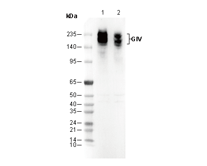 Lane 1: Mouse brain, Lane 2: Rat brain
IF
Validato da Selleck
-
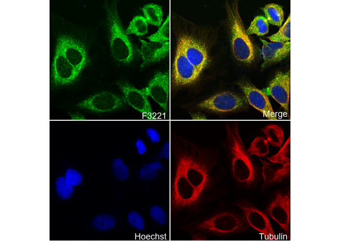 Immunofluorescent analysis of Hela cells using F3221 (green, 1:2000), Hoechst (blue) and tubulin (Red).