|
Come citare 1. Per la citazione nel testo (Materiali e Metodi): 2. Per la tabella delle risorse chiave: |
||
|
Numero verde: (877) 796-6397 -- Solo USA e Canada -- |
Fax: +1-832-582-8590 Ordini: +1-832-582-8158 |
Supporto tecnico: +1-832-582-8158 Ext:3 Si prega di fornire il numero dordine nelle-mail. Ci sforziamo di rispondere a tutte le richieste via e-mail entro un giorno lavorativo. |
Descrizione biologica
| Specificità | EB3 Antibody [J9A22] riconosce i livelli endogeni della proteina EB3 totale. |
|---|---|
| Contesto | EB3 (End-binding protein 3) è un membro cruciale della famiglia di proteine di tracciamento dell'estremità positiva dei microtubuli (+TIP), responsabile della regolazione della dinamica dei microtubuli e del mantenimento dell'architettura cellulare. EB3 presenta un dominio di omologia con la calponina (CH) che facilita il legame ai microtubuli e una regione C-terminale che consente interazioni con altre +TIP come CLASPs e Clip-170, influenzando così il comportamento dei microtubuli. La sua funzione principale prevede la stabilizzazione delle punte in crescita dei microtubuli riconoscendo i cappucci GTP/GDP-Pi e reclutando proteine regolatorie per garantire una corretta dinamica microtubulare. EB3 svolge ruoli significativi in vari processi cellulari, inclusa l'elongazione e la fusione dei mioblasti durante la miogenesi, dove organizza i microtubuli a livello del cortex cellulare per supportare la stabilità della protrusione della membrana. EB3 regola la segnalazione del calcio raggruppando i recettori dell'inositolo 1,4,5-trifosfato (IP3Rs), che controllano la permeabilità endoteliale, specialmente durante l'infiammazione. EB3 facilita la segnalazione del Ca²⁺ e l'interruzione della barriera dipendente dai microtubuli interagendo con gli IP3Rs tramite il motivo TxIP. EB3 influenza la morfologia del Golgi e l'organizzazione dei microtubuli non centrosomali ancorando i microtubuli decorati con CAMSAP2 all'apparato di Golgi. La rottura di EB3 porta a una compromissione della migrazione e dell'invasione cellulare. |
Informazioni sullutilizzo
| Applicazione | WB, IHC, IF, FCM | Diluizione |
|
||||||||
|---|---|---|---|---|---|---|---|---|---|---|---|
| Reattività | Human, Rat | ||||||||||
| Fonte | Rabbit Monoclonal Antibody | MW | 31 kDa | ||||||||
| Tampone di conservazione | PBS, pH 7.2+50% Glycerol+0.05% BSA+0.01% NaN3 | Conservazione (Dalla data di ricevimento) |
-20°C (avoid freeze-thaw cycles), 2 years | ||||||||
| WB |
Experimental Protocol:
Sample preparation
1. Tissue: Lyse the tissue sample by adding an appropriate volume of ice-cold RIPA/NP-40 Lysis Buffer (containing Protease Inhibitor Cocktail),and homogenize the tissue at a low temperature or lyse it by sonication on ice, then incubate on ice for 30 minutes. 2. Adherent cell: Aspirate the culture medium and transfer the cells into an EP tube. Wash the cells with ice-cold PBS twice. Add an appropriate volume of RIPA/NP-40 Lysis Buffer (containing Protease Inhibitor Cocktail), sonicate to lyse the cells, and incubate on ice for 30 minutes. 3. Suspension cell: Transfer the culture medium to a pre-cooled centrifuge tube. Centrifuge and aspirate the supernatant. Wash the cells with ice-cold PBS twice.Add an appropriate volume of RIPA/NP-40 Lysis Buffer (containing Protease Inhibitor Cocktail), sonicate to lyse the cells, and incubate on ice for 30 minutes. 4. Place the lysate into a pre-cooled microcentrifuge tube. Centrifuge at 4°C for 15 min. Collect the supernatant;
5. Remove a small volume of lysate to determine the protein concentration;
6. Combine the lysate with protein loading buffer. Boil 20 µL sample under 95-100°C for 5 min. Centrifuge for 5 min after cool down on ice.
Electrophoretic separation
1. According to the concentration of extracted protein, load appropriate amount of protein sample and marker onto SDS-PAGE gels for electrophoresis. Recommended separating gel (lower gel) concentration: 10%. Reference Table for Selecting SDS-PAGE Separation Gel Concentrations 2. Power up 80V for 30 minutes. Then the power supply is adjusted (110 V~150 V), the Marker is observed, and the electrophoresis can be stopped when the indicator band of the predyed protein Marker where the protein is located is properly separated. (Note that the current should not be too large when electrophoresis, too large current (more than 150 mA) will cause the temperature to rise, affecting the result of running glue. If high currents cannot be avoided, an ice bath can be used to cool the bath.)
Transfer membrane
1. Take out the converter, soak the clip and consumables in the pre-cooled converter;
2. Activate PVDF membrane with methanol for 1 min and rinse with transfer buffer;
3. Install it in the order of "black edge of clip - sponge - filter paper - filter paper - glue -PVDF membrane - filter paper - filter paper - sponge - white edge of clip"; 4. The protein was electrotransferred to PVDF membrane. ( 0.22 µm PVDF membrane is recommended )) Reference Table for Selecting PVDF Membrane Pore Size Specifications Recommended conditions for wet transfer: 200 mA, 60 min. ( Note that the transfer conditions can be adjusted according to the protein size. For high-molecular-weight proteins, a higher current and longer transfer time are recommended. However, ensure that the transfer tank remains at a low temperature to prevent gel melting.)
Block
1. After electrotransfer, wash the film with TBST at room temperature for 5 minutes;
2. Incubate the film in the blocking solution for 1 hour at room temperature;
3. Wash the film with TBST for 3 times, 5 minutes each time.
Antibody incubation
1. Use 5% skim milk powder to prepare the primary antibody working liquid (recommended dilution ratio for primary antibody 1:100000), gently shake and incubate with the film at 4°C overnight; 2. Wash the film with TBST 3 times, 5 minutes each time;
3. Add the secondary antibody to the blocking solution and incubate with the film gently at room temperature for 1 hour;
4. After incubation, wash the film with TBST 3 times for 5 minutes each time.
Antibody staining
1389. Add the prepared ECL luminescent substrate (or select other color developing substrate according to the second antibody) and mix evenly;
2. Incubate with the film for 1 minute, remove excess substrate (keep the film moist), wrap with plastic film, and expose in the imaging system.
|
Riferimenti
|
Dati di applicazione
WB
Validato da Selleck
-
 Lane 1: Human cerebellum
Lane 2: Human fetal brain
Lane 3: Rat muscle
Lane 4: SH-SY5Y
IF
Validato da Selleck
-
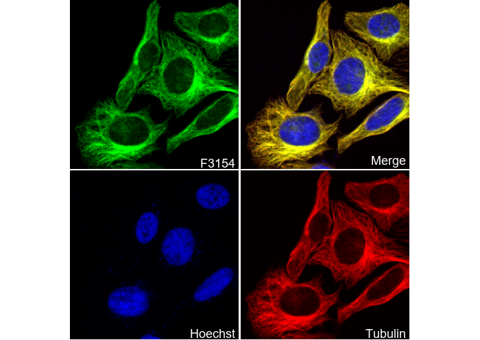 Immunofluorescent analysis of SH-SY5Y cells using F3154 (green, 1:250), Hoechst (blue) and tubulin (Red).