|
Come citare 1. Per la citazione nel testo (Materiali e Metodi): 2. Per la tabella delle risorse chiave: |
||
|
Numero verde: (877) 796-6397 -- Solo USA e Canada -- |
Fax: +1-832-582-8590 Ordini: +1-832-582-8158 |
Supporto tecnico: +1-832-582-8158 Ext:3 Si prega di fornire il numero dordine nelle-mail. Ci sforziamo di rispondere a tutte le richieste via e-mail entro un giorno lavorativo. |
Descrizione biologica
| Specificità | E4BP4/NFIL3 Antibody [J16L2] riconosce i livelli endogeni della proteina E4BP4/NFIL3 totale. |
|---|---|
| Contesto | Il fattore nucleare interleuchina 3 (NFIL3), noto anche come proteina legante E4 4 (E4BP4), è un repressore di numerosi geni. NFIL3 presenta un dominio a cerniera di leucina basica comprendente gli amminoacidi 73-146 all'interno dei suoi 462 residui; la parte N-terminale di questo dominio si lega direttamente al DNA, mentre la regione C-terminale facilita l'omodi o l'eterodimerizzazione della proteina. NFIL3 reprime i geni reclutando l'istone deacetilasi 2 e l'istone metiltransferasi G9a, regolando vari processi biologici, inclusi il ritmo circadiano, la vitalità cellulare e il metabolismo epatico. Nelle cellule immunitarie, NFIL3 è cruciale per lo switching di classe delle IgE delle cellule B e lo sviluppo delle cellule NK. Si lega al promotore Igε per stimolare la produzione di IgE. I topi deficienti di Nfil3 mostrano una significativa perdita di cellule NK a causa del ruolo del fattore nello sviluppo, nella maturazione e nella funzione delle cellule NK, e mostrano anche una elevata espressione di IL-12 p40 nel tessuto del colon, che porta alla differenziazione Th1 e alla colite spontanea. Le citochine Th2 sono influenzate in modo simile, con un'aumentata espressione di IL-5 e IL-13 nelle cellule Th2 Nfil3−/−. Inoltre, E4BP4/NFIL3 è essenziale per lo sviluppo delle cellule dendritiche CD8α+ convenzionali, poiché questo sottogruppo è assente nei topi E4BP4/NFIL3(-/-). |
Informazioni sullutilizzo
| Applicazione | WB, IP | Diluizione |
|
||||
|---|---|---|---|---|---|---|---|
| Reattività | Human, Mouse, Rat | ||||||
| Fonte | Rabbit Monoclonal Antibody | MW | 60 kda | ||||
| Tampone di conservazione | PBS, pH 7.2+50% Glycerol+0.05% BSA+0.01% NaN₃ | Conservazione (Dalla data di ricevimento) |
–20°C (avoid freeze-thaw cycles), 2 years | ||||
| WB |
Experimental Protocol:
Sample preparation
1. Tissue: Lyse the tissue sample by adding an appropriate volume of ice-cold RIPA/Nuclear Lysis Buffer (containing Protease Inhibitor Cocktail),and homogenize the tissue at a low temperature or lyse it by sonication on ice, then incubate on ice for 30 minutes. 2. Adherent cell: Aspirate the culture medium and transfer the cells into an EP tube. Wash the cells with ice-cold PBS twice. Add an appropriate volume of RIPA/Nuclear Lysis Buffer (containing Protease Inhibitor Cocktail), sonicate to lyse the cells, and incubate on ice for 30 minutes. 3. Suspension cell: Transfer the culture medium to a pre-cooled centrifuge tube. Centrifuge and aspirate the supernatant. Wash the cells with ice-cold PBS twice.Add an appropriate volume of RIPA/Nuclear Lysis Buffer (containing Protease Inhibitor Cocktail), sonicate to lyse the cells, and incubate on ice for 30 minutes. 4. Place the lysate into a pre-cooled microcentrifuge tube. Centrifuge at 4°C for 15 min. Collect the supernatant;
5. Remove a small volume of lysate to determine the protein concentration;
6. Combine the lysate with protein loading buffer. Boil 20 µL sample under 95-100°C for 5 min. Centrifuge for 5 min after cool down on ice.
Electrophoretic separation
1. According to the concentration of extracted protein, load appropriate amount of protein sample and marker onto SDS-PAGE gels for electrophoresis. Recommended separating gel (lower gel) concentration: 10%. Reference Table for Selecting SDS-PAGE Separation Gel Concentrations 2. Power up 80V for 30 minutes. Then the power supply is adjusted (110 V~150 V), the Marker is observed, and the electrophoresis can be stopped when the indicator band of the predyed protein Marker where the protein is located is properly separated. (Note that the current should not be too large when electrophoresis, too large current (more than 150 mA) will cause the temperature to rise, affecting the result of running glue. If high currents cannot be avoided, an ice bath can be used to cool the bath.)
Transfer membrane
1. Take out the converter, soak the clip and consumables in the pre-cooled converter;
2. Activate PVDF membrane with methanol for 1 min and rinse with transfer buffer;
3. Install it in the order of "black edge of clip - sponge - filter paper - filter paper - glue -PVDF membrane - filter paper - filter paper - sponge - white edge of clip"; 4. The protein was electrotransferred to PVDF membrane. ( 0.45 µm PVDF membrane is recommended ) Reference Table for Selecting PVDF Membrane Pore Size Specifications Recommended conditions for wet transfer: 200 mA, 120 min. ( Note that the transfer conditions can be adjusted according to the protein size. For high-molecular-weight proteins, a higher current and longer transfer time are recommended. However, ensure that the transfer tank remains at a low temperature to prevent gel melting.)
Block
1. After electrotransfer, wash the film with TBST at room temperature for 5 minutes;
2. Incubate the film in the blocking solution for 1 hour at room temperature;
3. Wash the film with TBST for 3 times, 5 minutes each time.
Antibody incubation
1. Use 5% skim milk powder to prepare the primary antibody working liquid (recommended dilution ratio for primary antibody 1:1000), gently shake and incubate with the film at 4°C overnight; 2. Wash the film with TBST 3 times, 5 minutes each time;
3. Add the secondary antibody to the blocking solution and incubate with the film gently at room temperature for 1 hour;
4. After incubation, wash the film with TBST 3 times for 5 minutes each time.
Antibody staining
696. Add the prepared ECL luminescent substrate (or select other color developing substrate according to the second antibody) and mix evenly;
2. Incubate with the film for 1 minute, remove excess substrate (keep the film moist), wrap with plastic film, and expose in the imaging system.
|
Riferimenti
|
Dati di applicazione
WB
Validato da Selleck
-
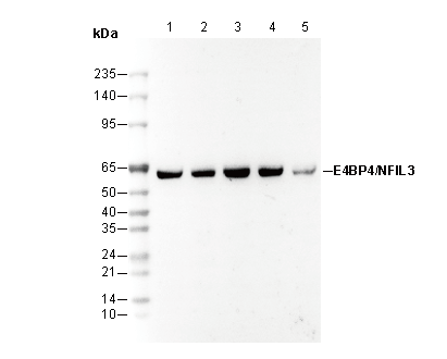 Lane 1: NK-92
Lane 2: Jurkat
Lane 3: Hep G2
Lane 4: HT-29
Lane 5: YB2/0