|
Come citare 1. Per la citazione nel testo (Materiali e Metodi): 2. Per la tabella delle risorse chiave: |
||
|
Numero verde: (877) 796-6397 -- Solo USA e Canada -- |
Fax: +1-832-582-8590 Ordini: +1-832-582-8158 |
Supporto tecnico: +1-832-582-8158 Ext:3 Si prega di fornire il numero dordine nelle-mail. Ci sforziamo di rispondere a tutte le richieste via e-mail entro un giorno lavorativo. |
Descrizione biologica
| Specificità | Collagen VII Antibody [K3G14] rileva i livelli endogeni della proteina totale di Collagen VII. |
|---|---|
| Contesto | Il collagene VII è un collagene fibrillare specializzato, principalmente responsabile della formazione delle fibrille di ancoraggio che stabilizzano la giunzione dermo-epidermica nella pelle. È codificato dal gene COL7A1 ed è espresso prevalentemente nei cheratinociti epidermici e nei fibroblasti dermici. Strutturalmente, il collagene VII è un omotrimero composto da tre catene pro-α1(VII), ciascuna caratterizzata da un dominio centrale a tripla elica (circa 1.530 amminoacidi) interrotto da 19 imperfezioni, inclusa una regione “cerniera” di 39 amminoacidi sensibile alle proteasi. Questo dominio centrale è affiancato da due domini non collagenici: NC-1 al N-terminale, che contiene moduli adesivi come ripetizioni di fibronectina di tipo III e un dominio A del fattore di von Willebrand, e NC-2 al C-terminale, che include un segmento simile a un inibitore di proteasi di Kunitz. Due molecole di collagene VII si assemblano in dimeri antiparalleli stabilizzati da legami disolfuro e si aggregano ulteriormente in fibrille di ancoraggio. Funzionalmente, il collagene VII ancora la membrana basale alla derma sottostante legandosi con alta affinità a componenti della membrana basale come la laminina-332 e il collagene IV, mantenendo così l'integrità della pelle; le sue mutazioni causano l'epidermolisi bollosa distrofica (EBD), una grave malattia cutanea vescicolare. |
Informazioni sullutilizzo
| Applicazione | IHC-Fr | Diluizione |
|
||
|---|---|---|---|---|---|
| Reattività | Human | ||||
| Fonte | Mouse Monoclonal Antibody | MW | |||
| Tampone di conservazione | PBS, pH 7.2+50% Glycerol+0.05% BSA+0.01% NaN3 | Conservazione (Dalla data di ricevimento) |
-20°C (avoid freeze-thaw cycles), 2 years | ||
| IHC |
Experimental Protocol:
Deparaffinization/Rehydration
1. Deparaffinize/hydrate sections:
2. Incubate sections in three washes of xylene for 5 min each.
3. Incubate sections in two washes of 100% ethanol for 10 min each.
4. Incubate sections in two washes of 95% ethanol for 10 min each.
5. Wash sections two times in dH2O for 5 min each.
6.Antigen retrieval: For Citrate: Heat slides in a microwave submersed in 1X citrate unmasking solution until boiling is initiated; continue with 10 min at a sub-boiling temperature (95°-98°C). Cool slides on bench top for 30 min.
Staining
1. Wash sections in dH2O three times for 5 min each.
2. Incubate sections in 3% hydrogen peroxide for 10 min.
3. Wash sections in dH2O two times for 5 min each.
4. Wash sections in wash buffer for 5 min.
5. Block each section with 100–400 µl of blocking solution for 1 hr at room temperature.
6. Remove blocking solution and add 100–400 µl primary antibody diluent in to each section. Incubate overnight at 4°C.
7. Remove antibody solution and wash sections with wash buffer three times for 5 min each.
8. Cover section with 1–3 drops HRPas needed. Incubate in a humidified chamber for 30 min at room temperature.
9. Wash sections three times with wash buffer for 5 min each.
10. Add DAB Chromogen Concentrate to DAB Diluent and mix well before use.
11. Apply 100–400 µl DAB to each section and monitor closely. 1–10 min generally provides an acceptable staining intensity.
12. Immerse slides in dH2O.
13. If desired, counterstain sections with hematoxylin.
14. Wash sections in dH2O two times for 5 min each.
15. Dehydrate sections: Incubate sections in 95% ethanol two times for 10 sec each; Repeat in 100% ethanol, incubating sections two times for 10 sec each; Repeat in xylene, incubating sections two times for 10 sec each.
16. Mount sections with coverslips and mounting medium.
|
Riferimenti
|
Dati di applicazione
IHC
Validato da Selleck
-
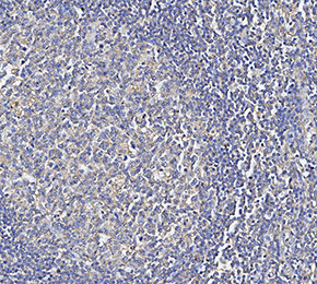 Immunohistochemical analysis of formalin fixed paraffin embedded human tonsils tissue with F3214 at 1:500 dilution.