|
Come citare 1. Per la citazione nel testo (Materiali e Metodi): 2. Per la tabella delle risorse chiave: |
||
|
Numero verde: (877) 796-6397 -- Solo USA e Canada -- |
Fax: +1-832-582-8590 Ordini: +1-832-582-8158 |
Supporto tecnico: +1-832-582-8158 Ext:3 Si prega di fornire il numero dordine nelle-mail. Ci sforziamo di rispondere a tutte le richieste via e-mail entro un giorno lavorativo. |
Descrizione biologica
| Specificità | CaV1.3 Antibody [G19D14] rileva i livelli endogeni della proteina CaV1.3 totale. |
|---|---|
| Contesto | L'accoppiamento eccitazione-contrazione cardiaca si riferisce alla sequenza di eventi in cui la stimolazione elettrica di un cardiomiocita innesca la contrazione muscolare nel cuore. I canali del Ca²⁺ di tipo L sono fondamentali per questo processo, in quanto facilitano l'influsso di calcio e contribuiscono all'eccitabilità della membrana. Sono stati identificati quattro sottotipi di canali del Ca²⁺ di tipo L: Cav1.1, Cav1.2, Cav1.3 e Cav1.4. Cav1.1 si trova prevalentemente nel muscolo scheletrico, mentre Cav1.4 è espresso principalmente nella retina e in alcune cellule immunitarie. Cav1.3 è presente nel cuore, nelle regioni somatodendritiche dei neuroni, nelle cellule endocrine e nelle cellule sensoriali. Nel cuore, l'attività di Cav1.3 è modulata da più neurotrasmettitori. La fosforilazione da parte della proteina chinasi A (PKA) dipendente dal cAMP avviene ai residui di serina 1743 e 1816 all'interno della regione C-terminale. La proteina chinasi C (PKC) regola anche Cav1.3 in modo isoenzima-specifico tramite fosforilazione alla serina 81 nel dominio N-terminale. Inoltre, lo splicing alternativo all'interno del C-terminale influenza il comportamento del canale, diminuendo in particolare l'inattivazione Ca²⁺-dipendente. Funzionalmente, Cav1.3 contribuisce al pacemaker cardiaco e alla conduzione atrioventricolare (AV). La disfunzione di Cav1.3 è stata associata ad anomalie del nodo senoatriale e del nodo AV, nonché allo sviluppo di fibrillazione atriale. |
Informazioni sullutilizzo
| Applicazione | IHC, FCM | Diluizione |
|
||
|---|---|---|---|---|---|
| Reattività | Human, Mouse | ||||
| Fonte | Mouse Monoclonal Antibody | MW | |||
| Tampone di conservazione | PBS, pH 7.2+50% Glycerol+0.05% BSA+0.01% NaN3 | Conservazione (Dalla data di ricevimento) |
-20°C (avoid freeze-thaw cycles), 2 years | ||
| IHC |
Experimental Protocol:
Deparaffinization/Rehydration
1. Deparaffinize/hydrate sections:
2. Incubate sections in three washes of xylene for 5 min each.
3. Incubate sections in two washes of 100% ethanol for 10 min each.
4. Incubate sections in two washes of 95% ethanol for 10 min each.
5. Wash sections two times in dH2O for 5 min each.
6.Antigen retrieval: For Citrate: Heat slides in a microwave submersed in 1X citrate unmasking solution until boiling is initiated; continue with 10 min at a sub-boiling temperature (95°-98°C). Cool slides on bench top for 30 min.
Staining
1. Wash sections in dH2O three times for 5 min each.
2. Incubate sections in 3% hydrogen peroxide for 10 min.
3. Wash sections in dH2O two times for 5 min each.
4. Wash sections in wash buffer for 5 min.
5. Block each section with 100–400 µl of blocking solution for 1 hr at room temperature.
6. Remove blocking solution and add 100–400 µl primary antibody diluent in to each section. Incubate overnight at 4°C.
7. Remove antibody solution and wash sections with wash buffer three times for 5 min each.
8. Cover section with 1–3 drops HRPas needed. Incubate in a humidified chamber for 30 min at room temperature.
9. Wash sections three times with wash buffer for 5 min each.
10. Add DAB Chromogen Concentrate to DAB Diluent and mix well before use.
11. Apply 100–400 µl DAB to each section and monitor closely. 1–10 min generally provides an acceptable staining intensity.
12. Immerse slides in dH2O.
13. If desired, counterstain sections with hematoxylin.
14. Wash sections in dH2O two times for 5 min each.
15. Dehydrate sections: Incubate sections in 95% ethanol two times for 10 sec each; Repeat in 100% ethanol, incubating sections two times for 10 sec each; Repeat in xylene, incubating sections two times for 10 sec each.
16. Mount sections with coverslips and mounting medium.
|
Riferimenti
|
Dati di applicazione
IHC
Validato da Selleck
-
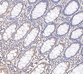 Immunohistochemical analysis of formalin fixed paraffin embedded human colorectal cancer tissue with F3798 at 1:1000 dilution.