|
Come citare 1. Per la citazione nel testo (Materiali e Metodi): 2. Per la tabella delle risorse chiave: |
||
|
Numero verde: (877) 796-6397 -- Solo USA e Canada -- |
Fax: +1-832-582-8590 Ordini: +1-832-582-8158 |
Supporto tecnico: +1-832-582-8158 Ext:3 Si prega di fornire il numero dordine nelle-mail. Ci sforziamo di rispondere a tutte le richieste via e-mail entro un giorno lavorativo. |
Descrizione biologica
| Specificità | CA19-9 Antibody [C17A13] rileva i livelli endogeni della proteina CA 19-9. |
|---|---|
| Contesto | Il CA19-9 (antigene carboidrato 19-9), noto anche come Sialyl Lewis A (sLeᵃ), è un antigene del gruppo sanguigno Lewis sialilato, un epitopo glicanico tetrasaccaride composto da acido sialico, galattosio, N-acetilglucosamina e fucosio, biosintetizzato tramite la via del gruppo sanguigno Lewis e che richiede un antigene Lewis funzionale per la sua formazione. Il CA19-9 è espresso su glicoproteine e glicolipidi delle membrane delle cellule epiteliali, in particolare nel pancreas, nel tratto biliare, nello stomaco e nel colon, e può essere rilasciato nel flusso sanguigno. È l'attuale biomarcatore sierico gold standard per l'adenocarcinoma pancreatico. Funzionalmente, il CA19-9 serve come biomarcatore (per diagnosi, prognosi, monitoraggio della risposta al trattamento e rilevamento delle recidive), un predittore (correlato con il carico tumorale, lo stadio e la resecabilità) e un promotore della progressione del cancro facilitando l'adesione mediata dalla E-selectina, migliorando l'angiogenesi, modulando le risposte immunitarie e influenzando le interazioni del microambiente tumorale. Oltre al cancro al pancreas, il CA19-9 è elevato in altre neoplasie gastrointestinali e in alcune malattie benigne, ed è oggetto di indagine come bersaglio terapeutico tramite anticorpi, vaccini, inibitori della biosintesi e sistemi di somministrazione di farmaci guidati dal CA19-9. |
Informazioni sullutilizzo
| Applicazione | IHC, FCM, ELISA | Diluizione |
|
||
|---|---|---|---|---|---|
| Reattività | Human | ||||
| Fonte | Mouse Monoclonal Antibody | MW | |||
| Tampone di conservazione | PBS, pH 7.2+50% Glycerol+0.05% BSA+0.01% NaN3 | Conservazione (Dalla data di ricevimento) |
-20°C (avoid freeze-thaw cycles), 2 years | ||
| IHC |
Experimental Protocol:
Deparaffinization/Rehydration
1. Deparaffinize/hydrate sections:
2. Incubate sections in three washes of xylene for 5 min each.
3. Incubate sections in two washes of 100% ethanol for 10 min each.
4. Incubate sections in two washes of 95% ethanol for 10 min each.
5. Wash sections two times in dH2O for 5 min each.
6.Antigen retrieval: For Citrate: Heat slides in a microwave submersed in 1X citrate unmasking solution until boiling is initiated; continue with 10 min at a sub-boiling temperature (95°-98°C). Cool slides on bench top for 30 min.
Staining
1. Wash sections in dH2O three times for 5 min each.
2. Incubate sections in 3% hydrogen peroxide for 10 min.
3. Wash sections in dH2O two times for 5 min each.
4. Wash sections in wash buffer for 5 min.
5. Block each section with 100–400 µl of blocking solution for 1 hr at room temperature.
6. Remove blocking solution and add 100–400 µl primary antibody diluent in to each section. Incubate overnight at 4°C.
7. Remove antibody solution and wash sections with wash buffer three times for 5 min each.
8. Cover section with 1–3 drops HRPas needed. Incubate in a humidified chamber for 30 min at room temperature.
9. Wash sections three times with wash buffer for 5 min each.
10. Add DAB Chromogen Concentrate to DAB Diluent and mix well before use.
11. Apply 100–400 µl DAB to each section and monitor closely. 1–10 min generally provides an acceptable staining intensity.
12. Immerse slides in dH2O.
13. If desired, counterstain sections with hematoxylin.
14. Wash sections in dH2O two times for 5 min each.
15. Dehydrate sections: Incubate sections in 95% ethanol two times for 10 sec each; Repeat in 100% ethanol, incubating sections two times for 10 sec each; Repeat in xylene, incubating sections two times for 10 sec each.
16. Mount sections with coverslips and mounting medium.
|
Riferimenti
|
Dati di applicazione
IHC
Validato da Selleck
-
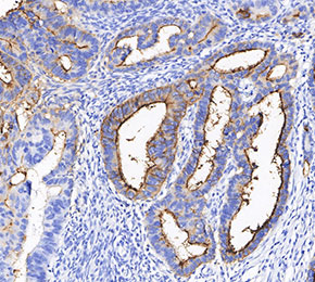 Immunohistochemical analysis of formalin fixed paraffin embedded human colorectal cancer tissue with F2466 at 1:100 dilution.