|
Come citare 1. Per la citazione nel testo (Materiali e Metodi): 2. Per la tabella delle risorse chiave: |
||
|
Numero verde: (877) 796-6397 -- Solo USA e Canada -- |
Fax: +1-832-582-8590 Ordini: +1-832-582-8158 |
Supporto tecnico: +1-832-582-8158 Ext:3 Si prega di fornire il numero dordine nelle-mail. Ci sforziamo di rispondere a tutte le richieste via e-mail entro un giorno lavorativo. |
Descrizione biologica
| Specificità | ATG4B Antibody [P14M3] rileva i livelli endogeni della proteina ATG4B totale. |
|---|---|
| Contesto | ATG4B è una cruciale cisteina proteasi nell'Autophagy, responsabile dell'elaborazione e del riciclo delle proteine della famiglia ATG8 come LC3 e GABARAP, consentendo così la biogenesi degli autofagosomi negli eucarioti. ATG4B contiene una triade catalitica composta da Cys74, Asp278 e His280 all'interno di un ripiegamento simile alla papaina, con un anello regolatore e Trp142 che mantengono l'enzima in uno stato autoinibito fino a quando l'interazione con il substrato induce cambiamenti conformazionali per l'idrolisi del legame peptidico. I motivi N-terminali migliorano ulteriormente la specificità per LC3 coniugato alla fosfatidiletanolamina (PE) sulle membrane autofagiche. La funzione primaria di ATG4B è quella di esporre la glicina C-terminale sulla pro-LC3 attraverso una scissione di priming iniziale, preparandola per la coniugazione alla PE tramite la cascata ubiquitina-simile ATG7/ATG3, e successivamente di catalizzare la delipidazione di LC3-PE sulla membrana autofagosomica esterna, facilitando il riciclo di LC3 libero. Con la più ampia selettività del substrato tra i paraloghi ATG4, ATG4B favorisce principalmente LC3, bilanciando l'allungamento della membrana autofagosomica, l'ancoraggio e il flusso autofagico. La sua attività è strettamente regolata attraverso modificazioni redox a Cys292 e Cys361 e dalla fosforilazione, adattando l'Autophagy allo stress cellulare; la sovraespressione di ATG4B wild-type può bloccare la lipidazione di LC3 per eccessiva deconiugazione, mentre mutanti cataliticamente inattivi come C74A sequestrano LC3 e inibiscono l'Autophagy. La disregolazione dell'attività di ATG4B è legata alla tumorigenesi a causa di un flusso autofagico compromesso. |
Informazioni sullutilizzo
| Applicazione | WB, FCM | Diluizione |
|
||||
|---|---|---|---|---|---|---|---|
| Reattività | Mouse, Human | ||||||
| Fonte | Rabbit Monoclonal Antibody | MW | 44 kDa | ||||
| Tampone di conservazione | PBS, pH 7.2+50% Glycerol+0.05% BSA+0.01% NaN3 | Conservazione (Dalla data di ricevimento) |
-20°C (avoid freeze-thaw cycles), 2 years | ||||
| WB |
Experimental Protocol:
Sample preparation
1. Tissue: Lyse the tissue sample by adding an appropriate volume of ice-cold RIPA/NP-40 Lysis Buffer (containing Protease Inhibitor Cocktail),and homogenize the tissue at a low temperature. 2. Adherent cell: Aspirate the culture medium and wash the cells with ice-cold PBS twice. Lyse the cells by adding an appropriate volume of RIPA/NP-40 Lysis Buffer (containing Protease Inhibitor Cocktail) and put the sample on ice for 5 min. 3. Suspension cell: Transfer the culture medium to a pre-cooled centrifuge tube. Centrifuge and aspirate the supernatant. Wash the cells with ice-cold PBS twice. Lyse the cells by adding an appropriate volume of RIPA/NP-40 Lysis Buffer (containing Protease Inhibitor Cocktail) and put the sample on ice for 5 min. 4. Place the lysate into a pre-cooled microcentrifuge tube. Centrifuge at 4°C for 15 min. Collect the supernatant;
5. Remove a small volume of lysate to determine the protein concentration;
6. Combine the lysate with protein loading buffer. Boil 20 µL sample under 95-100°C for 5 min. Centrifuge for 5 min after cool down on ice.
Electrophoretic separation
1. According to the concentration of extracted protein, load appropriate amount of protein sample and marker onto SDS-PAGE gels for electrophoresis. Recommended separating gel (lower gel) concentration: 10%. Reference Table for Selecting SDS-PAGE Separation Gel Concentrations 2. Power up 80V for 30 minutes. Then the power supply is adjusted (110 V~150 V), the Marker is observed, and the electrophoresis can be stopped when the indicator band of the predyed protein Marker where the protein is located is properly separated. (Note that the current should not be too large when electrophoresis, too large current (more than 150 mA) will cause the temperature to rise, affecting the result of running glue. If high currents cannot be avoided, an ice bath can be used to cool the bath.)
Transfer membrane
1. Take out the converter, soak the clip and consumables in the pre-cooled converter;
2. Activate PVDF membrane with methanol for 1 min and rinse with transfer buffer;
3. Install it in the order of "black edge of clip - sponge - filter paper - filter paper - glue -PVDF membrane - filter paper - filter paper - sponge - white edge of clip"; 4. The protein was electrotransferred to PVDF membrane. ( 0.45 µm PVDF membrane is recommended ) Reference Table for Selecting PVDF Membrane Pore Size Specifications Recommended conditions for wet transfer: 200 mA, 120 min. ( Note that the transfer conditions can be adjusted according to the protein size. For high-molecular-weight proteins, a higher current and longer transfer time are recommended. However, ensure that the transfer tank remains at a low temperature to prevent gel melting.)
Block
1. After electrotransfer, wash the film with TBST at room temperature for 5 minutes;
2. Incubate the film in the blocking solution for 1 hour at room temperature;
3. Wash the film with TBST for 3 times, 5 minutes each time.
Antibody incubation
1. Use 5% skim milk powder to prepare the primary antibody working liquid (recommended dilution ratio for primary antibody 1:1000), gently shake and incubate with the film at 4°C overnight; 2. Wash the film with TBST 3 times, 5 minutes each time;
3. Add the secondary antibody to the blocking solution and incubate with the film gently at room temperature for 1 hour;
4. After incubation, wash the film with TBST 3 times for 5 minutes each time.
Antibody staining
1. Add the prepared ECL luminescent substrate (or select other color developing substrate according to the second antibody) and mix evenly;
2. Incubate with the film for 1 minute, remove excess substrate (keep the film moist), wrap with plastic film, and expose in the imaging system.
|
Riferimenti
|
Dati di applicazione
WB
Validato da Selleck
-
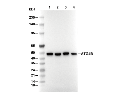 Lane 1: Ramos, Lane 2: 293T, Lane 3: Jurkat, Lane 4: Hela