|
Come citare 1. Per la citazione nel testo (Materiali e Metodi): 2. Per la tabella delle risorse chiave: |
||
|
Numero verde: (877) 796-6397 -- Solo USA e Canada -- |
Fax: +1-832-582-8590 Ordini: +1-832-582-8158 |
Supporto tecnico: +1-832-582-8158 Ext:3 Si prega di fornire il numero dordine nelle-mail. Ci sforziamo di rispondere a tutte le richieste via e-mail entro un giorno lavorativo. |
Descrizione biologica
| Specificità | Artemis Antibody [H20J21] rileva i livelli endogeni della proteina Artemis totale. |
|---|---|
| Contesto | Artemis (DCLRE1C, noto anche come SNM1C) è una nucleasi nucleare espressa ubiquitariamente appartenente alla superfamiglia delle metallo-β-lattamasi (MBL), essenziale per la riparazione del DNA tramite giunzione di estremità non omologhe (NHEJ) e la ricombinazione V(D)J nei linfociti in via di sviluppo. Artemis è una proteina di 692 amminoacidi con un dominio catalitico N-terminale (residui 1-360) che comprende un ripiegamento MBL centrale (sandwich α/β-β/α) e un sottodominio β-CASP incorporato contenente i motivi conservati 1-4 (His33, His35, His115, Asp116 che coordinano gli ioni Zn²⁺ M1/M2) e i motivi A-C per l'elaborazione degli acidi nucleici, oltre a una coda regolatoria C-terminale in gran parte non strutturata (residui 361-692) che auto-inibisce l'attività e media il legame di DNA-PKcs. DNA-PKcs fosforila Artemis (ad esempio, a Thr127, Ser251) al riconoscimento dei DSB, attivando la sua attività endonucleasica struttura-specifica per aprire le estremità codificanti sigillate a forcina nella ricombinazione V(D)J, tagliare i "overhang" 5'/3', i "flap" e le bolle nella NHEJ per la ligazione XRCC4/LIG4, e processare le estremità complesse indotte da IR antagonizzando l'HR tramite l'interazione 53BP1-PTIP. ATM/ATR fosforilano ulteriormente Artemis per il recupero del checkpoint G2/M e della fase S, promuovendo l'attivazione di Cdk1-ciclina B e la degradazione della ciclina E tramite SCF^{Fbw7}. Le mutazioni bialleliche causano immunodeficienza combinata grave radiosensibile (RS-SCID) con difetti di giunzione V(D)J e instabilità genomica che predispongono alla malignità. |
Informazioni sullutilizzo
| Applicazione | WB, IP | Diluizione |
|
||||
|---|---|---|---|---|---|---|---|
| Reattività | Human, Monkey | ||||||
| Fonte | Rabbit Monoclonal Antibody | MW | 90 kDa | ||||
| Tampone di conservazione | PBS, pH 7.2+50% Glycerol+0.05% BSA+0.01% NaN3 | Conservazione (Dalla data di ricevimento) |
-20°C (avoid freeze-thaw cycles), 2 years | ||||
| WB |
Experimental Protocol:
Sample preparation
1. Tissue: Lyse the tissue sample by adding an appropriate volume of ice-cold RIPA/Nuclear Lysis Buffer (containing Protease Inhibitor Cocktail),and homogenize the tissue at a low temperature. 2. Adherent cell: Aspirate the culture medium and wash the cells with ice-cold PBS twice. Lyse the cells by adding an appropriate volume of RIPA/Nuclear Lysis Buffer (containing Protease Inhibitor Cocktail) and put the sample on ice for 5 min. 3. Suspension cell: Transfer the culture medium to a pre-cooled centrifuge tube. Centrifuge and aspirate the supernatant. Wash the cells with ice-cold PBS twice. Lyse the cells by adding an appropriate volume of RIPA/Nuclear Lysis Buffer (containing Protease Inhibitor Cocktail) and put the sample on ice for 5 min. 6. Add protein loading buffer, heat 20 μL of the sample at 95~100°C for 5 min, let it cool down on ice and then centrifuge for 5 min. Electrophoretic separation
1. According to the concentration of extracted protein, load appropriate amount of protein sample and marker onto SDS-PAGE gels for electrophoresis. Recommended separating gel (lower gel) concentration: 10%. Reference Table for Selecting SDS-PAGE Separation Gel Concentrations 2. Power up 80V for 30 minutes. Then the power supply is adjusted (110 V~150 V), the Marker is observed, and the electrophoresis can be stopped when the indicator band of the predyed protein Marker where the protein is located is properly separated. (Note that the current should not be too large when electrophoresis, too large current (more than 150 mA) will cause the temperature to rise, affecting the result of running glue. If high currents cannot be avoided, an ice bath can be used to cool the bath.)
Transfer membrane
1. Take out the converter, soak the clip and consumables in the pre-cooled converter;
2. Activate PVDF membrane with methanol for 1 min and rinse with transfer buffer;
3. Install it in the order of "black edge of clip - sponge - filter paper - filter paper - glue -PVDF membrane - filter paper - filter paper - sponge - white edge of clip"; 4. The protein was electrotransferred to PVDF membrane. ( 0.45 µm PVDF membrane is recommended ) Reference Table for Selecting PVDF Membrane Pore Size Specifications Recommended conditions for wet transfer: 200 mA, 120 min. ( Note that the transfer conditions can be adjusted according to the protein size. For high-molecular-weight proteins, a higher current and longer transfer time are recommended. However, ensure that the transfer tank remains at a low temperature to prevent gel melting.)
Block
1. After electrotransfer, wash the film with TBST at room temperature for 5 minutes;
2. Incubate the film in the blocking solution for 1 hour at room temperature;
3. Wash the film with TBST for 3 times, 5 minutes each time.
Antibody incubation
1. Use 5% skim milk powder to prepare the primary antibody working liquid (recommended dilution ratio for primary antibody 1:1000), gently shake and incubate with the film at 4°C overnight; 2. Wash the film with TBST 3 times, 5 minutes each time;
3. Add the secondary antibody to the blocking solution and incubate with the film gently at room temperature for 1 hour;
4. After incubation, wash the film with TBST 3 times for 5 minutes each time.
Antibody staining
1. Add the prepared ECL luminescent substrate (or select other color developing substrate according to the second antibody) and mix evenly;
2. Incubate with the film for 1 minute, remove excess substrate (keep the film moist), wrap with plastic film, and expose in the imaging system. (Exposure time of at least 60s is recommended)
|
Riferimenti
|
Dati di applicazione
WB
Validato da Selleck
-
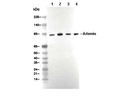 Lane 1: Jurkat, Lane 2: HT-29, Lane 3: PC-3, Lane 4: MCF-7