|
Come citare 1. Per la citazione nel testo (Materiali e Metodi): 2. Per la tabella delle risorse chiave: |
||
|
Numero verde: (877) 796-6397 -- Solo USA e Canada -- |
Fax: +1-832-582-8590 Ordini: +1-832-582-8158 |
Supporto tecnico: +1-832-582-8158 Ext:3 Si prega di fornire il numero dordine nelle-mail. Ci sforziamo di rispondere a tutte le richieste via e-mail entro un giorno lavorativo. |
Descrizione biologica
| Specificità | 8-Hydroxy-2'-deoxyguanosine Antibody [K11E8] rileva sia l'8-idrossi-2'-deossiguanosina (8-OH-dG) endogena che esogena. |
|---|---|
| Contesto | La 8-idrossi-2'-deossiguanosina (8-OHdG) è un 2'-deossiribonucleoside purinico modificato formato dall'idrossilazione della base guaninica in posizione C8 nel DNA, tipicamente derivante dal danno ossidativo causato dalle specie reattive dell'ossigeno (ROS). Strutturalmente, consiste in una molecola di deossiguanosina con un gruppo idrossilico sul carbonio 8 dell'anello guaninico. L'8-OHdG è ampiamente riconosciuta come un biomarcatore chiave dello stress ossidativo a causa della sua elevata prevalenza tra le lesioni ossidative del DNA. Si accumula sia nel DNA nucleare che mitocondriale e può essere misurata in vari campioni biologici, inclusi urina, tessuti e DNA leucocitario. Funzionalmente, l'8-OHdG serve come indicatore di danno al DNA endogeno ed è stata ampiamente utilizzata per valutare il rischio e la progressione di cancro, invecchiamento e malattie degenerative. La sua presenza riflette l'esposizione cellulare ad agenti ossidativi ed è fondamentale per valutare l'impatto dei fattori ambientali e dello stile di vita sulla stabilità genomica. |
Informazioni sullutilizzo
| Applicazione | IHC-P | Diluizione |
|
||
|---|---|---|---|---|---|
| Reattività | Species independent | ||||
| Fonte | Mouse Monoclonal Antibody | MW | |||
| Tampone di conservazione | PBS, pH 7.2+50% Glycerol+0.05% BSA+0.01% NaN3 | Conservazione (Dalla data di ricevimento) |
-20°C (avoid freeze-thaw cycles), 2 years | ||
| IHC |
Experimental Protocol:
Deparaffinization/Rehydration
1. Deparaffinize/hydrate sections:
2. Incubate sections in three washes of xylene for 5 min each.
3. Incubate sections in two washes of 100% ethanol for 10 min each.
4. Incubate sections in two washes of 95% ethanol for 10 min each.
5. Wash sections two times in dH2O for 5 min each.
6.Antigen retrieval: For Citrate: Heat slides in a microwave submersed in 1X citrate unmasking solution until boiling is initiated; continue with 10 min at a sub-boiling temperature (95°-98°C). Cool slides on bench top for 30 min.
Staining
1. Wash sections in dH2O three times for 5 min each.
2. Incubate sections in 3% hydrogen peroxide for 10 min.
3. Wash sections in dH2O two times for 5 min each.
4. Wash sections in wash buffer for 5 min.
5. Block each section with 100–400 µl of blocking solution for 1 hr at room temperature.
6. Remove blocking solution and add 100–400 µl primary antibody diluent in to each section. Incubate overnight at 4°C.
7. Remove antibody solution and wash sections with wash buffer three times for 5 min each.
8. Cover section with 1–3 drops HRPas needed. Incubate in a humidified chamber for 30 min at room temperature.
9. Wash sections three times with wash buffer for 5 min each.
10. Add DAB Chromogen Concentrate to DAB Diluent and mix well before use.
11. Apply 100–400 µl DAB to each section and monitor closely. 1–10 min generally provides an acceptable staining intensity.
12. Immerse slides in dH2O.
13. If desired, counterstain sections with hematoxylin.
14. Wash sections in dH2O two times for 5 min each.
15. Dehydrate sections: Incubate sections in 95% ethanol two times for 10 sec each; Repeat in 100% ethanol, incubating sections two times for 10 sec each; Repeat in xylene, incubating sections two times for 10 sec each.
16. Mount sections with coverslips and mounting medium.
|
Riferimenti
|
Dati di applicazione
IHC
Validato da Selleck
-
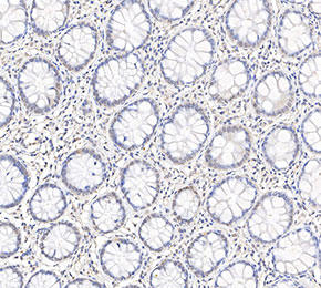 Immunohistochemical analysis of formalin fixed paraffin embedded human colorectal cancer tissue with F1714 at 1:50 dilution.