|
Come citare 1. Per la citazione nel testo (Materiali e Metodi): 2. Per la tabella delle risorse chiave: |
||
|
Numero verde: (877) 796-6397 -- Solo USA e Canada -- |
Fax: +1-832-582-8590 Ordini: +1-832-582-8158 |
Supporto tecnico: +1-832-582-8158 Ext:3 Si prega di fornire il numero dordine nelle-mail. Ci sforziamo di rispondere a tutte le richieste via e-mail entro un giorno lavorativo. |
Descrizione biologica
| Specificità | 53BP1 Antibody [M24M17] rileva i livelli endogeni della proteina 53BP1 totale. |
|---|---|
| Contesto | La 53BP1 (p53-binding protein 1), codificata dal gene TP53BP1 sul cromosoma 15, è una proteina chiave di risposta al DNA Damage/DNA Repair che regola la riparazione delle rotture a doppio filamento (DSB), in particolare attraverso il percorso di giunzione delle estremità non omologhe (NHEJ). Strutturalmente, è una proteina scaffold, caratterizzata da siti di fosforilazione N-terminali, una Minimal Focus Forming Region (FFR) per la localizzazione della cromatina, domini Tudor in tandem che riconoscono H4K20me2, e domini BRCT che interagiscono con p53 e γH2AX. Funzionalmente, la 53BP1 stabilizza le estremità del DNA, facilita la loro ligazione e influenza l'equilibrio tra NHEJ e ricombinazione omologa (HR), prevenendo un'eccessiva resezione del DNA. È ampiamente espressa in vari tessuti, specialmente nelle cellule a rapida divisione, e i suoi livelli aumentano in seguito a DNA Damage/DNA Repair. Clinicamente, la 53BP1 è cruciale nella biologia del cancro, dove la sua deficienza influisce sulla sensibilità agli inibitori di PARP (PARPi) nei tumori con deficienza di BRCA1. Serve come biomarcatore per l'efficienza della DNA Damage/DNA Repair, la prognosi del cancro e la risposta alla terapia, rendendola un bersaglio promettente per interventi terapeutici che manipolano le vie di DNA Damage/DNA Repair. |
Informazioni sullutilizzo
| Applicazione | WB, IHC, IF, FCM | Diluizione |
|
||||||||
|---|---|---|---|---|---|---|---|---|---|---|---|
| Reattività | Human, Mouse, Rat | ||||||||||
| Fonte | Rabbit Monoclonal Antibody | MW | 214 kDa | ||||||||
| Tampone di conservazione | PBS, pH 7.2+50% Glycerol+0.05% BSA+0.01% NaN3 | Conservazione (Dalla data di ricevimento) |
-20°C (avoid freeze-thaw cycles), 2 years | ||||||||
| WB |
Experimental Protocol:
Sample preparation
1. Tissue: Lyse the tissue sample by adding an appropriate volume of ice-cold RIPA/NP-40 Lysis Buffer (containing Protease Inhibitor Cocktail),and homogenize the tissue at a low temperature or lyse it by sonication on ice, then incubate on ice for 30 minutes. 2. Adherent cell: Aspirate the culture medium and transfer the cells into an EP tube. Wash the cells with ice-cold PBS twice. Add an appropriate volume of RIPA/NP-40 Lysis Buffer (containing Protease Inhibitor Cocktail), sonicate to lyse the cells, and incubate on ice for 30 minutes. 3. Suspension cell: Transfer the culture medium to a pre-cooled centrifuge tube. Centrifuge and aspirate the supernatant. Wash the cells with ice-cold PBS twice.Add an appropriate volume of RIPA/NP-40 Lysis Buffer (containing Protease Inhibitor Cocktail), sonicate to lyse the cells, and incubate on ice for 30 minutes. 4. Place the lysate into a pre-cooled microcentrifuge tube. Centrifuge at 4°C for 15 min. Collect the supernatant;
5. Remove a small volume of lysate to determine the protein concentration;
6. Combine the lysate with protein loading buffer. Boil 20 µL sample under 95-100°C for 5 min. Centrifuge for 5 min after cool down on ice.
Electrophoretic separation
1. According to the concentration of extracted protein, load appropriate amount of protein sample and marker onto SDS-PAGE gels for electrophoresis. Recommended separating gel (lower gel) concentration: 5%. Reference Table for Selecting SDS-PAGE Separation Gel Concentrations 2. Power up 80V for 30 minutes. Then the power supply is adjusted (110 V~150 V), the Marker is observed, and the electrophoresis can be stopped when the indicator band of the predyed protein Marker where the protein is located is properly separated. (Note that the current should not be too large when electrophoresis, too large current (more than 150 mA) will cause the temperature to rise, affecting the result of running glue. If high currents cannot be avoided, an ice bath can be used to cool the bath.)
Transfer membrane
1. Take out the converter, soak the clip and consumables in the pre-cooled converter;
2. Activate PVDF membrane with methanol for 1 min and rinse with transfer buffer;
3. Install it in the order of "black edge of clip - sponge - filter paper - filter paper - glue -PVDF membrane - filter paper - filter paper - sponge - white edge of clip"; 4. The protein was electrotransferred to PVDF membrane. ( 0.45 µm PVDF membrane is recommended ) Reference Table for Selecting PVDF Membrane Pore Size Specifications Recommended conditions for wet transfer: 250 mA, 180 min. ( Note that the transfer conditions can be adjusted according to the protein size. For high-molecular-weight proteins, a higher current and longer transfer time are recommended. However, ensure that the transfer tank remains at a low temperature to prevent gel melting.)
Block
1. After electrotransfer, wash the film with TBST at room temperature for 5 minutes;
2. Incubate the film in the blocking solution for 1 hour at room temperature;
3. Wash the film with TBST for 3 times, 5 minutes each time.
Antibody incubation
1. Use 5% skim milk powder to prepare the primary antibody working liquid (recommended dilution ratio for primary antibody 1:1000), gently shake and incubate with the film at 4°C overnight; 2. Wash the film with TBST 3 times, 5 minutes each time;
3. Add the secondary antibody to the blocking solution and incubate with the film gently at room temperature for 1 hour;
4. After incubation, wash the film with TBST 3 times for 5 minutes each time.
Antibody staining
1389. Add the prepared ECL luminescent substrate (or select other color developing substrate according to the second antibody) and mix evenly;
2. Incubate with the film for 1 minute, remove excess substrate (keep the film moist), wrap with plastic film, and expose in the imaging system.
|
Riferimenti
|
Dati di applicazione
WB
Validato da Selleck
-
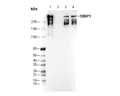 Lane 1: HAP1
Lane 2: HAP1 (KO 53BP1)
Lane 3: HeLa
Lane 4: HepG2
IF
Validato da Selleck
-
 Immunofluorescent analysis of HepG2 cells using F2158 (green, 1:100), Hoechst (blue) and tubulin (Red).