
solo per uso di ricerca
Quinolinic acid NMDAR Agonista
N. Cat.: S3624
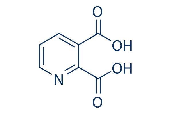
Struttura chimica
Peso molecolare: 167.12
Controllo Qualità (Quality Control)
- Citato in Nature Medicine per la sua qualità superiore
- COA
- HPLC
- SDS
- Scheda tecnica
Informazioni chimiche, conservazione e stabilità (Chemical Information, Storage & Stability)
| Peso molecolare | 167.12 | Formula | C7H5NO4 |
Conservazione (Dalla data di ricezione) | |
|---|---|---|---|---|---|
| N. CAS | 89-00-9 | Scarica SDF | Conservazione delle soluzioni stock |
|
|
| Sinonimi | pyridine-2,3-dicarboxylic acid, QUIN | Smiles | C1=CC(=C(N=C1)C(=O)O)C(=O)O | ||
Solubilità (Solubility)
|
In vitro |
DMSO
: 25 mg/mL
(149.59 mM)
Water : 3 mg/mL Ethanol : Insoluble |
Calcolatore di Molarità
|
In vivo |
|||||
Calcolatore di formulazione in vivo (Soluzione chiara)
Passo 1: Inserire le informazioni di seguito (Consigliato: Un animale aggiuntivo per tenere conto della perdita durante l'esperimento)
Passo 2: Inserire la formulazione in vivo (Questo è solo il calcolatore, non la formulazione. Contattateci prima se non c'è una formulazione in vivo nella sezione Solubilità.)
Risultati del calcolo:
Concentrazione di lavoro: mg/ml;
Metodo per preparare il liquido master di DMSO: mg farmaco predissolto in μL DMSO ( Concentrazione del liquido master mg/mL, Vi preghiamo di contattarci prima se la concentrazione supera la solubilità del DMSO del lotto del farmaco. )
Metodo per preparare la formulazione in vivo: Prendere μL DMSO liquido master, quindi aggiungereμL PEG300, mescolare e chiarire, quindi aggiungereμL Tween 80, mescolare e chiarire, quindi aggiungere μL ddH2O, mescolare e chiarire.
Metodo per preparare la formulazione in vivo: Prendere μL DMSO liquido master, quindi aggiungere μL Olio di mais, mescolare e chiarire.
Nota: 1. Si prega di assicurarsi che il liquido sia limpido prima di aggiungere il solvente successivo.
2. Assicurarsi di aggiungere il/i solvente/i in ordine. È necessario assicurarsi che la soluzione ottenuta, nell'aggiunta precedente, sia una soluzione limpida prima di procedere all'aggiunta del solvente successivo. Metodi fisici come il vortex, gli ultrasuoni o il bagno d'acqua calda possono essere utilizzati per facilitare la dissoluzione.
Meccanismo d'azione (Mechanism of Action)
| Targets/IC50/Ki |
NMDAR
|
|---|---|
| In vitro |
QUIN is an agonist of N-methyl-D-aspartate (NMDA) receptor, and it has a high in vivo potency as an excitotoxin. Although QUIN has an uptake system, its neuronal degradation enzyme is rapidly saturated, and the rest of extracellular QUIN can continue stimulating the NMDA receptor. QUIN (10 μM) prevents of glutamate-induced excitotoxicity in primary cultures of rat cerebellar granule neurons, nevertheless mature organotypic cultures of rat corticostriatal system or caudate nucleus chronically exposed to 100 nM QUIN for up to 7 weeks show focal degeneration characterized by the presence of vacuoles in neuropil, swollen dendrites, occasional swollen post-synaptic elements, and degenerated neurons. In vitro QUIN treatment of human primary foetal neurons leads to a substantial increase of tau phosphorylation at multiple positions. The increase in QUIN-induced phosphorylation of tau is attributed to a decrease in the expression and activity of the major tau phosphatases. QUIN can inhibit B monoamine oxidase (MAO-B) in human brain synaptosomal mitochondria and also can be a potent inhibitor of phosphoenolpyruvate carboxykinase (EC 4.1.1.32) from rat liver cytoplasm, an important enzyme in the gluconeogenesis pathway that converts oxaloacetate to phosphoenolpyruvate. QUIN can increase free radical production by inducing NOS activity in astrocytes and neurons, leading to oxidative stress, increasing both poly(ADP-ribose) polymerase (PARP) activity and extracellular lactate dehydrogenase (LDH) activity.
|
| In vivo |
Quinolinic acid (QUIN), a neuroactive metabolite of the kynurenine pathway, is normally presented in nanomolar concentrations in human brain and cerebrospinal fluid (CSF) and is often implicated in the pathogenesis of a variety of human neurological diseases. The concentration of QUIN varies among different brain regions, with the cerebral cortex containing approximately 1.8 nmol/g wet weight; almost 2-fold than that found in the hippocampus (1 nmol/g wet weight). Intraarterial administration of either micromolar or millimolar concentrations of QUIN results in only negligible accumulations of this metabolite in the brain, suggesting that the central nervous system (CNS) appears to be well protected by the blood brain barrier (BBB) from peripheral QUIN. QUIN can also increase glutamate release and inhibit its reuptake by astrocytes, thus increasing its concentration in the microenvironments, causing neurotoxicity and also limiting glutamate to glutamine recycling in astrocytes by decreasing glutamine synthetase activity. Intrastriatal injection of QUIN provokes a decrease of cellular respiration and ATP levels.
|
Riferimenti |
|
Informazioni sullo studio clinico (Clinical Trial Information)
(dati da https://clinicaltrials.gov, aggiornato il 2024-05-22)
| Numero NCT | Reclutamento | Condizioni | Sponsor/Collaboratori | Data di inizio | Fasi |
|---|---|---|---|---|---|
| NCT05513807 | Not yet recruiting | Delayed Graft Function |
Assistance Publique - Hôpitaux de Paris|Institut National de la Santé Et de la Recherche Médicale France|Centre National de la Recherche Scientifique France |
2022-11 | Phase 3 |
| NCT05718310 | Recruiting | Migraine|Migraine Disorders|Pain |
IRCCS National Neurological Institute C. Mondino Foundation|University of Pavia |
2022-11-01 | Not Applicable |
| NCT05969990 | Recruiting | Migraine in Children |
Qilu Hospital of Shandong University |
2022-01-01 | -- |
| NCT04342975 | Completed | AKI |
Mayo Clinic|Elysium Health |
2020-12-01 | Phase 2 |

I prodotti sono solo per uso di ricerca. Non per uso umano. Non vendiamo a pazienti.
©Copyright 2013 Selleck Chemicals. Tutti i diritti riservati.